
Clavicular Notch · What Is It, Location, Purpose, and More

Published: Feb 04, 2025
Author: Anna Hernández, MD
Editor: Alyssa Haag
Editor: Ian Mannarino, MD, MBA
Editor: Kelsey LaFayette, DNP, ARNP, FNP-C
Illustrator: Jessica Reynolds, MS
Copyeditor: David G. Walker
7-day free trial

Go deeper with Osmosis

Osmosis is a learning platform with videos, questions, and AI tools to help you master topics like this.

4.8 · 12,000+ reviews
Watch quick, visual videos
Practice with Qbank-style questions
Use AI to explain, quiz, and review
Study anytime with the mobile app
Start free trial

No credit card · Cancel anytime

What is the clavicular notch?

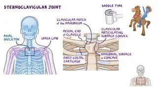
The clavicular notch is a small, oval-shaped depression located on either side of the sternum. It is angled posteriorly, laterally, and superiorly and articulates with the sternal end of each clavicle to form the sternoclavicular joints (the only bony articulation that attaches the upper limb to the axial skeleton).

Learn deeper with Osmosis

Master this topic faster with videos, questions, and AI.

Used by 8M+ healthcare learners.

Start free trial

No credit card · Cancel anytime

Where is the clavicular notch?

The clavicular notch is found laterally on either side of the manubrium of the sternum. The sternum is the flat, elongated bone that sits in the middle of the anterior part of the thoracic cage. The manubrium (which is the trapezoid bone that forms the superior portion of the sternum) has a shallow, U-shaped depression called the jugular or suprasternal notch that can be easily palpated at the base of the neck. The jugular notch is deepened by the sternal ends of the clavicles, which bulge out from the clavicular notches in the manubrium. Inferior and lateral to the clavicular notches is the first sternocostal joint, which is the site of attachment of the first costal cartilage to the manubrium of the sternum.

What is the purpose of the clavicular notch?

The clavicular notch articulates with the medial or sternal end of each clavicle to form the two sternoclavicular joints. The sternoclavicular joint is a saddle type of synovial joint, named so because the manubrial articulating surface is concave and the clavicular articulating surface is convex. The two articulating surfaces fit together the same way that a cowboy sits on a horse’s saddle, hence the name saddle joint. 

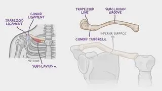
The sternoclavicular joint has an articular disc that is firmly attached to the fibrous joint capsule by the sternoclavicular ligaments and the interclavicular ligament. The ligaments surrounding the fibrous capsule are the anterior and the posterior sternoclavicular ligaments, which reinforce the joint capsule anteriorly and posteriorly; the interclavicular ligament, which strengthens the sternoclavicular joint superiorly; and the costoclavicular ligament, which anchors the inferior surface of the medial end of the clavicle to the first costal cartilage. The costoclavicular ligament also provides stability to the sternoclavicular joint by limiting the elevation of the pectoral girdle.

The main function of the sternoclavicular joint is to support the range of motion of the upper limb, as well as provide a stable connection between the upper limbs and the upper body.
Students say Osmosis is 100% worth it

Because Osmosis saves them time. Lowers stress. And actually helps them remember when it counts.

I used Osmosis to prepare for my first medical school licensing exam! Super helpful and interactive for people who may not do great with just pages of text info!

Cecilia Ruiz

Cecilia Ruiz

MD student

Sayan Misra

I have used Osmosis for about four years. Best thing I have ever used for my medical studies.

Sayan Misra

Sayan Misra

Med student

Osmosis videos are superior because they define simple concepts, tell a story with a clear progression, and provide context.

Jay Pate

Jay Pate

Dental student

References


Altalib, A. A., Miao, K. H., & Menezes, R. G. (2022). Anatomy, Thorax, Sternum. In StatPearls. Retrieved from https://www.ncbi.nlm.nih.gov/books/NBK541141/#:~:text=The%20sternum%20is%20a%20partially,forming%20the%20anterior%20rib%20cage


Hansen, J. T., & Netter, F. H. (Eds.). (2014). Netter’s clinical anatomy (4th edition). Elsevier.


Harrison, L. (2019). The thoracic cage. In Anatomical Basis of Injury. Retrieved from https://uhlibraries.pressbooks.pub/atpanatomy/chapter/7-4-the-thoracic-cage/


Standring, S., & Gray, H. (Eds.). (2021). Gray’s anatomy: The anatomical basis of clinical practice (42nd ed.). Elsevier.