Chapters:

Introduction0:00–0:20

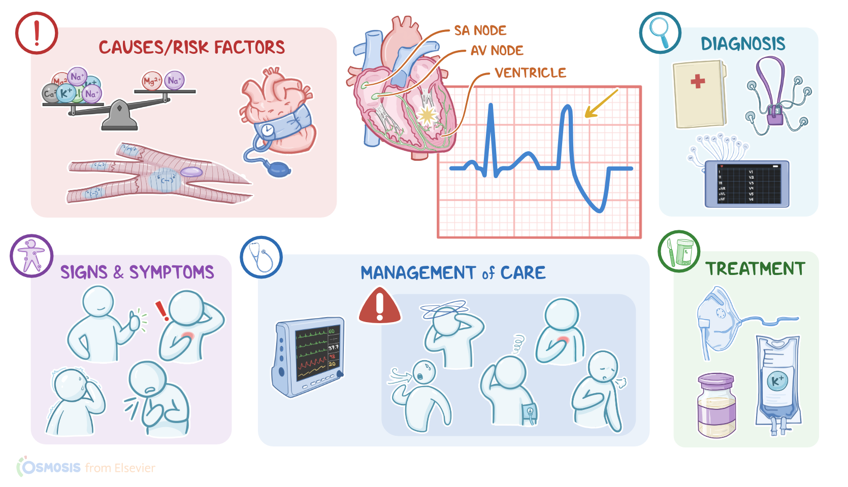
Premature ventricular contractions, or simply PVCs, are a type of arrhythmia, where an extra beat starts in a point in the ventricles, causing the ventricles to contract earlier than normal in the cardiac cycle.
Now let’s look at the normal electrical conduction pathway in the heart on an ECG, which shows how the depolarization wave flows through the heart during each heartbeat.

Physiology0:20–2:29

The normal electrical activity of the heart starts in the sinoatrial or SA node, which is considered the pacemaker of the heart.
Then, the impulse is conducted through the atrium, creating the P wave on an ECG. And when the atrial muscle cells get depolarized, they contract, pushing blood from the atria into the ventricles.
From the atrium, electrical activity goes to the atrioventricular, or AV node, where the impulse propagation speed slows way down; this is the PR interval on an ECG.
This pause allows the atria to contract while the ventricles fill with blood. From the AV node, the depolarization wave goes through the Bundle of His, then the right and left branches of the Bundle, and finally through the Purkinje fibers, which deliver the current to the right and left ventricles, causing them to depolarize.
This triggers simultaneous contraction of both ventricles, pushing blood into the systemic and pulmonary circulations, and it’s represented by the QRS complex on an ECG.
Finally, the ventricles repolarize to prepare for the next cycle, which allows them to relax and fill with blood, called diastole.
And on ECG, ventricular repolarization will create a T wave, while the pause between ventricular depolarization and repolarization is represented by the ST segment.
Sometimes, immediately after the T wave, there’s a U wave, which represents late repolarization of the ventricles. Now, the 3 main causes of premature ventricular contractions are enhanced automaticity, triggered activity, and reentry.

Causes & risk factors2:29–4:51

Now, automaticity is the property of cells in the SA node, AV node, Bundle of His, and the Purkinje fibers to generate an electrical impulse, so they’re all pacemakers but follow the SA node’s lead.
However, when there’s a stressor that irritates the pacemaker cells in the ventricles, it could enhance their automaticity and cause them to fire early.
Risk factors can include electrolyte imbalance like hypokalemia and hypomagnesemia, damage to cardiac tissue like ischemia, and anything that increases sympathetic activity like hyperthyroidism, anxiety, or substances like caffeine or methamphetamine.
Next, there’s triggered activity where a previous impulse causes an additional depolarization in the ventricle called an “after-depolarization” and this can occur in a pattern, like once after every other beat.
The cause of it is not fully understood but it’s likely due to dysfunctional ion channels or injury to cardiac tissue. A final type of ventricular ectopic focus is a reentrant loop, where a depolarization wave encounters tissue that doesn’t depolarize, which can be something like scar tissue after a myocardial infarction, and as a result the wave starts going around and around that tissue - forming what’s called a reentrant loop.
On the other hand, non-modifiable risk factors include being assigned male at birth, black race, and advanced age. Now, regardless of the cause of the premature ventricular contraction it will lead to a pathological process that disrupts the normal heart rhythm.

Pathology4:51–6:07

This depends on when the ectopic PVC occurs. So, the impulse from the ectopic beat typically travels back up to the atrium and this is called a retrograde conduction.
If the ectopic beat happens right after the atrium has just depolarized, the cells in the conduction pathway are in its refractory period, so the impulse doesn’t make it pass the AV node, so there’s no extra atrial or ventricular contraction.
When the SA node fires again, the heart returns to sinus rhythm. On the other hand, if the ectopic beat occurs later, then the atrium could already be out of its refractory period, so the retrograde conduction will cause an extra atrial contraction.
Furthermore, when the impulse reaches the SA node it’ll cause a depolarization after the atrium’s already contracted, so the impulse doesn’t go anywhere and it essentially resets the node.
Clients with premature ventricular contractions are typically asymptomatic, but some clients present with palpitations, diminished or absent peripheral pulses, in addition to feeling that the heart skips a beat, which is typically followed by a fluttering sensation.

Clinical manifestations6:07–6:33

Other signs and symptoms include anxiety, lightheadedness, dyspnea, and chest pain.The diagnosis of premature ventricular contractions starts with the client's history and physical assessment, followed by cardiac monitoring using an electrocardiogram or a Holter monitor, which is basically a portable ECG that records for a 24-hour period.

Diagnosis6:33–8:47

So, on the ECG, the heart rate may vary depending on the intrinsic rate and the number of PVCs, and the rhythm can be irregular.
Keep in mind P waves are rarely seen right before a premature ventricular contraction and, as a consequence, PR intervals can’t be measured.
Sometimes, P waves are seen after a PVC, which is caused by retrograde conduction of the ectopic impulse to the atria. The QRS complex is typically wide and distorted, meaning that it lasts for more than 0.12 seconds.
On the other hand, T waves are typically larger and are opposite to the direction of the QRS complex. On ECG, premature ventricular contractions are grouped based on their shape into unifocal and multifocal.
Unifocal means that the premature ventricular contractions are similar in shape, so these contractions arise from the same ectopic focus.
On the other hand, multifocal means that the contractions have different shapes, so they arise from different ectopic foci.
Premature ventricular contractions can also be grouped based on their number into couplets, meaning that there are two consecutive PVCs; or triplets, meaning that there are three consecutive PVCs.
Frequency can also be used to group PVCs into bigeminy, meaning that the PVC happens every other beat; or trigeminy, meaning that the PVC happens every third beat.
Typically, three or more consecutive PVCs means that there’s ventricular tachycardia. All right, now, no treatment is needed for asymptomatic clients with premature ventricular contractions.

Treatment8:47–9:37

In contrast, in symptomatic clients, treatment is typically geared at identifying and eliminating the underlying causes.
For example, oxygen therapy is used in clients with hypoxia, and electrolyte replacements are prescribed in clients with electrolyte imbalances.
Certain medications can also be administered, such as beta-blockers, lidocaine, and amiodarone. Finally, clients with refractory cases may require radiofrequency ablation, which is a surgical procedure in which small scars are made to the part of the heart that forms the abnormal signals.All right, let’s look at the nursing care you’ll be providing for a client with PVCs.

Management and care9:37–10:30

Your priority goal is to monitor for complications. Start by initiating continuous cardiac monitoring to assess their heart rhythm, and monitoring for complications by assessing vital signs, and checking for symptoms of decreased cardiac output.
Immediately report three or more consecutive PVCs, lightheadedness, hypotension, chest pain, tachypnea, or shortness of breath to the healthcare provider.
Administer supplemental oxygen, and the prescribed medications, as indicated. Then, assist the healthcare team in identifying the underlying cause of the arrhythmia.
Now, on to client and family teaching. Begin by explaining that a PVC is a common type of abnormal heart rhythm where there are extra heartbeats that are less effective in pumping oxygenated blood around the body.

General client and family teaching10:30–12:25

If your client is prescribed a medication, teach them about how the medication helps maintain a more normal heart rhythm, and instruct them to take each medication exactly as directed.
Then, talk to your client about lifestyle modifications that can help keep their heart healthy. Encourage them to maintain a healthy weight, and to engage in moderate physical exercise regularly, as prescribed by their healthcare provider.
Instruct them to eat plenty of fruits, vegetables, and whole grains, as well as lean meats, and low-fat or fat-free dairy products.
Provide them with a list of foods they can select from, and teach them how to choose foods that are low in sodium. Also review the importance of avoiding triggers such as alcohol and caffeine, as well as any products that contain nicotine or tobacco, including cigarettes, e-cigarettes, and chewing tobacco.
If they need help quitting smoking, provide them with counseling and refer them to resources for support. Also teach them about the importance of adequate sleep, review relaxation techniques, and talk to them about strategies they can use to deal with stressful situations.
Finally, teach them how to take their pulse and blood pressure at home, and instruct them to let their healthcare provider know immediately if there are any significant changes in their heart rate or blood pressure; or if they experience fatigue, exercise intolerance, shortness of breath, or chest pain to their healthcare provider.

Review12:25–14:07

All right, as a quick recap…. Premature ventricular contractions are a type of arrhythmia, where an extra beat starts in the ventricle.
This causes the ventricles to contract earlier than normal.The 3 main causes of premature ventricular contractions are enhanced automaticity, triggered activity, and reentry, and risk factors can be modifiable, such as electrolyte imbalances, underlying cardiovascular disease and anything that increases sympathetic activity, like hyperthyroidism, anxiety or substances like caffeine or methamphetamine; or non-modifiable, such as being assigned male at birth, black race, and advanced age.
Most clients with PVCs are asymptomatic, but symptoms can include a fluttering sensation, palpitations, and diminished or absent peripheral pulses.
PVCs are usually diagnosed with cardiac monitoring, such as an EKG or holter. Treatment is generally not needed unless the client is symptomatic or has an underlying cause.
In these cases, treatment can include oxygen for hypoxia, electrolyte replacement, or medications such as beta blockers, lidocaine, or amiodarone.
Goals of nursing care are to monitor for complications. Education for client’s with PVCs should center on general health promotion, home care, and when to contact the healthcare provider.