Epidural and subdural hematoma: Nursing
Intracranial hemorrhage or ICH for short, refers to bleeding inside the skull. Types of intracranial hemorrhage include epidural hematoma, subdural hematoma, subarachnoid hemorrhage, and intracerebral bleeding. Now, epidural hematoma is the collection of blood in the epidural space, which is the space between the dura mater and inner surface of the skull. In contrast, subdural hematoma is the collection of blood in the subdural space, meaning between the dura mater and the arachnoid mater.
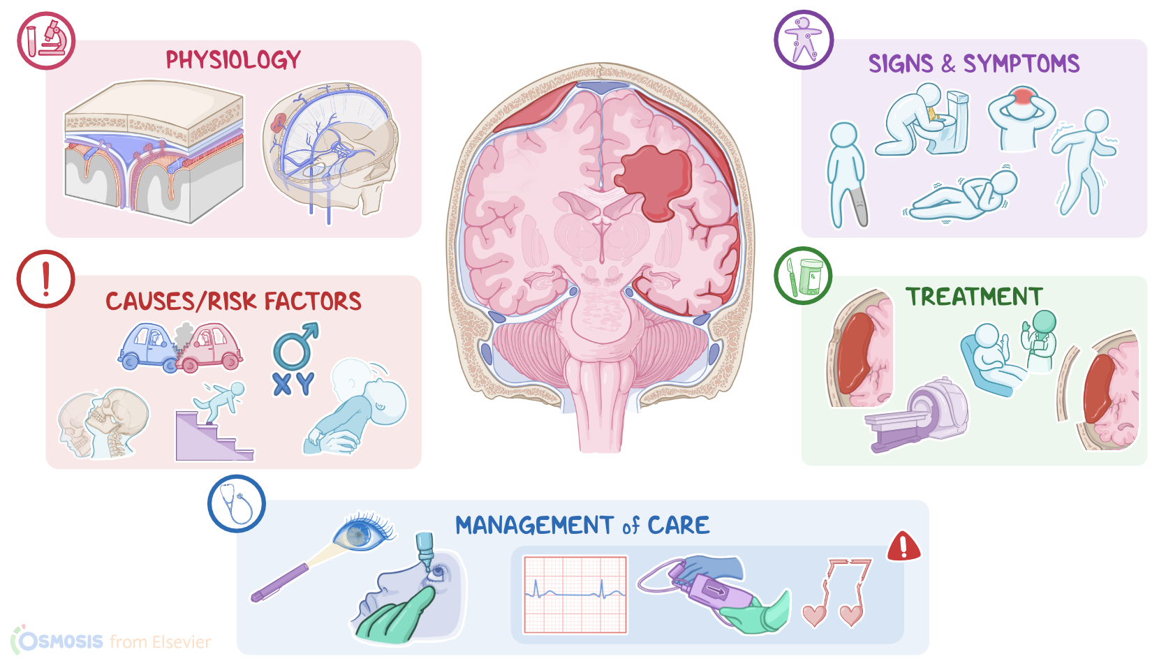
Let’s start by looking at the physiology of the meninges. This is a membrane that wraps around the brain and protects it from the outside environment. It’s made up of three meningeal layers. From outside to inside, these are the dura mater, arachnoid mater, and pia mater. Between the arachnoid and pia mater there’s the subarachnoid space, which houses the cerebrospinal fluid. The cerebrospinal fluid is a clear, watery liquid that cushions the brain from impact and bathes it in nutrients.
Now, the dura mater consists of an external and an internal layer. The external layer of the dura mater adheres tightly to the inner surface of the skull, so the epidural space is only a virtual space in the cranium, meaning the space isn’t appreciable unless pathology is present.
Between the external layer of the dura mater and the inner surface of the skull, there are meningeal arteries that supply meninges. The internal layer of the dura mater lies above the arachnoid mater, the two are separated by the subdural space. The subdural space plays a major role in venous blood drainage in the brain. The dura mater also forms dural venous sinuses which carry the venous blood from the brain. These sinuses are connected to the veins outside of the cranium by bridging veins.
| EPIDURAL AND SUBDURAL HEMATOMA | ||
| KEY POINTS | NOTES | |
| DEFINITION |
| |
| PHYSIOLOGY |
| |
| CAUSES AND RISK FACTORS |
| |
| PATHOPHYSIOLOGY |
| |
| SIGNS AND SYMPTOMS |
| |
| DIAGNOSIS |
| |
| TREATMENT |
| |
| MANAGEMENT OF CARE |
| |
| PATIENT AND FAMILY TEACHING |
| |
No notes for this video yet
Try adding a note below