Cholelithiasis · What Is It, Causes, Treatment, and More

Published: Oct 21, 2025
Author: Ali Syed, PharmD
Editor: Alyssa Haag, MD
Editor: Józia McGowan, DO
Editor: Kelsey LaFayette, DNP, ARNP, FNP-C
Editor: Anna Hernández, MD
Illustrator: Jillian Dunbar
Copyeditor: David G. Walker
7-day free trial

Go deeper with Osmosis

Osmosis is a learning platform with videos, questions, and AI tools to help you master topics like this.

4.8 · 12,000+ reviews
Watch quick, visual videos
Practice with Qbank-style questions
Use AI to explain, quiz, and review
Study anytime with the mobile app
Start free trial

No credit card · Cancel anytime

What is cholelithiasis?

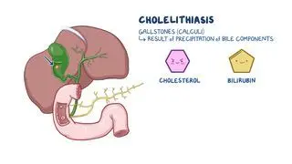
Cholelithiasis refers to the formation of gallstones, which are hardened deposits of digestive fluid that form in the gallbladder. The gallbladder is a small, pear-shaped organ that lies beneath the liver and its main job is to store bile made by the liver. Bile is a digestive fluid made of cholesterol, bile salts, and bilirubin and gets released into the small intestine through the cystic duct and common bile duct to aid in fat digestion.  

Gallstones can range in size and an individual may develop several small gallstones, one large gallstone, or a combination of different sized gallstones. In the United States, roughly 6% of males and 9% of females have cholelithiasis, most of whom are completely asymptomatic.  

Learn deeper with Osmosis

Master this topic faster with videos, questions, and AI.

Used by 8M+ healthcare learners.

Start free trial

No credit card · Cancel anytime

How do you pronounce cholelithiasis?

Cholelithiasis is pronounced as kow-luh-luh-thai-uh-suhs. 

What causes cholelithiasis?

The exact cause of cholelithiasis is not entirely clear, although it’s often the result of multiple factors. Cholelithiasis most commonly results from a chemical imbalance within the contents of the gallbladder. This occurs either when there is too much cholesterol or bilirubin in the bile, or when there are not enough bile salts and acids or phospholipids to dissolve it. The two most common types of gallstones are cholesterol gallstones and pigment gallstones 

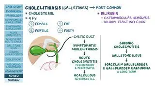
Cholesterol gallstones are the most common type, and they develop when the amount of cholesterol excreted by the liver exceeds what bile salts can dissolve, leading to the precipitation of cholesterol into yellow cholesterol stones. Risk factors associated with the development of cholesterol gallstones include high cholesterol blood levels, high fat and low fiber diets, obesity, sedentary living, and rapid weight loss. Female sex, as well as pregnancy and oral contraceptive pills, are associated with higher estrogen levels, which increase the activity of HMG-CoA reductase, the enzyme responsible for cholesterol synthesis in the liver, also making them risk factors for gallstones. Finally, increased incidence of cholesterol gallstones is found among certain ethnicities, particularly Indigenous people of North America and Europeans. 

Bilirubin gallstones are less common and are formed when bilirubin in the bile combines with calcium to form the solid precipitate calcium bilirubinate. Bilirubin is a chemical that’s produced when the body breaks down red blood cells and is excreted through the liver. Increased bilirubin can result from certain conditions, such as liver cirrhosisbiliary tract infections; cystic fibrosis; blood disorders, such as sickle cell anemia or leukemia; and gastrointestinal diseases associated with impaired bile reabsorption, such as Crohn disease.  

In general, gallstone disease may also be caused by concentrated bile, which may occur when the gallbladder is not emptying correctly or frequently enough due to low motility or contraction, increasing the risk of gallstones. 

What are the signs and symptoms of cholelithiasis?

Most individuals with cholelithiasis are asymptomatic, as gallstones typically do not cause any symptoms unless they obstruct the cystic duct, bile ducts, or both. Risk of obstruction varies depending on the size and location of the gallstone. Small gallstones are more likely to exit the gallbladder and block the cystic or bile ducts, while larger gallstones are more likely to stay in the gallbladder and not cause any symptoms. 

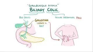
When symptoms occur, the most common manifestation are gallbladder attacks, or biliary colic, characterized by severe, rapidly intensifying abdominal pain localized to the upper right or central abdomen. Abdominal pain associated with biliary colic may radiate to the upper back, between the shoulder blades and to the right shoulder, and may last anywhere from 15 minutes to several hours. The episode typically subsides when the gallstone dislodges, relieving the pain. Other abdominal symptoms, such as clay-colored stools, bloating, indigestion, belching, and gas, may also occur. 

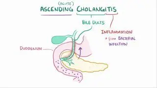
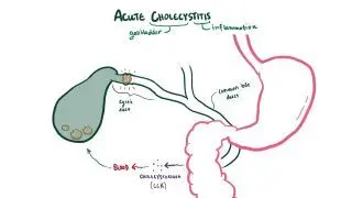
Persistent blockage of the cystic duct may lead to increased pressure within the gallbladder, leading to more severe, long-lasting pain and even acute cholecystitis, which is an inflammation of the gallbladder. Alternatively, gallstones that pass the cystic duct and block the common bile duct may result in choledocholithiasis, which is when a gallstone becomes lodged in the choledoco, also known as the common bile duct. Choledocholithiasis can cause itchy skin and/or yellowing of the skin and eyes, also known as jaundice, due to the accumulation of bilirubin. Finally, persistent blockage of the bile ducts may also result in acute pancreatitis, a serious condition characterized by high fever, sweating, chills, rapid heartbeat, nausea, vomiting, and diarrhea. 

How is cholelithiasis diagnosed?

Cholelithiasis  is diagnosed based on a review of symptoms, medical history, and physical examination. In most cases, an abdominal ultrasound may be performed to look for the presence of gallstones within the gallbladder as a first-line test. This technique is painless and non-invasive, so it’s often the preferred imaging technique. However, very small stones (<3 mm) may not produce the characteristic acoustic shadowing, making them more difficult to detect. Additionally, if the individual hasn’t eaten in a while, the gallbladder may be collapsed, making stones more difficult to see. Sometimes, overlying intestinal gas or fatty tissue can also obscure the gallbladder, limiting its visualization.  

Endoscopic retrograde cholangiopancreatography (ERCP) is a diagnostic and potential treatment procedure that involves the insertion of a long, thin, and flexible tube with a camera attached (i.e., an endoscope) through the mouth, down toward the small intestine. During an ERCP, an imaging dye is injected into the body to highlight the ducts of the biliary system. If gallstones are present in the bile ducts, they may be removed by the endoscope; however, the endoscope cannot remove stones inside the gallbladder.   

Magnetic resonance cholangiopancreatography (MRCP) is another diagnostic procedure in which the bile ducts are examined with magnetic resonance imaging. This procedure provides a more detailed image than the abdominal ultrasound, is less invasive than ERCP, and is a safer alternative for most individuals. In urgent cases (e.g., ascending cholangitis), ERCP may be done before MRCP. In an endoscopic ultrasound (EUS), an endoscope is passed through the mouth, down toward the common bile duct and gallbladder region, to screen for gallstones not visible in an abdominal ultrasound. An ERCP may follow MRCP or EUS in order to remove the visualized gallstones from the bile ducts.   

Other imaging tests that may be used to diagnose cholelithiasis include oral cholecystography, a hepatobiliary iminodiacetic acid (HIDA) scan, and CT scan. In addition, blood samples may be drawn to screen for infections, jaundice, or pancreatitis. 
How is cholelithiasis treated?

Treatment options for cholelithiasis depend on the age of the individual, overall health, medical history, and severity of symptoms, as well as the size, location, and quantity of gallstones. 

In asymptomatic individuals, cholelithiasis may not require any treatment besides observation. Symptomatic cholelithiasis is often resolved with gallbladder removal surgery, known as cholecystectomy. This eliminates the risk for future recurrences and allows bile to flow unobstructed from the liver to the small intestine.   

Nowadays, laparoscopic cholecystectomy is the most commonly performed technique as it is minimally invasive and involves the use of a laparoscope, or a narrow tube with a camera. The laparoscope is inserted through one of several small incisions in the abdomen to visualize and remove the gallbladder. If a laparoscopic cholecystectomy is not successful or if an individual experiences complications, an open cholecystectomy may be performed. Unlike laparoscopic cholecystectomy, open surgery involves one large incision to the abdomen and a longer healing period.  

In situations where cholecystectomy is not a viable option due to infection of the biliary tree, a percutaneous drainage may be put in place until a cholecystectomy can be performed. After a cholecystectomy, certain individuals may require a course of antibiotics, such as ciprofloxacin and metronidazole, to prevent or treat residual infection. 

Does cholelithiasis require surgery?

Cholelithiasis only requires surgery (i.e., cholecystectomy) if it causes significant symptoms or complications. Many people with gallstones remain asymptomatic and do not need treatment.  

What are the side effects of gallbladder removal surgery?

Gallbladder removal surgery, or cholecystectomy, is generally a safe procedure, but it can lead to certain side effects due to changes in bile flow and digestion. The gallbladder’s main function is to store and concentrate bile. Without it, bile constantly flows into the small intestine instead of being stored and released in response to meals. In some cases, this can result in abdominal discomfort, nausea, bloating, and diarrhea, especially when eating fatty foods.  

These symptoms usually improve within a few months of surgery; however, dietary measures are generally recommended to improve digestion. Management involves eating smaller and frequent meals, limiting high-fat foods (e.g., fried foods, cheese, butter, cream, and processed snacks), increasing fiber intake, and staying well-hydrated. 

What are the most important facts to know about cholelithiasis?

Cholelithiasis, or gallstones, are hardened deposits of digestive fluid that form in the gallbladder. Cholelithiasis commonly results from a chemical imbalance within the contents of the gallbladder in which the bile contains excessive cholesterol or bilirubin. The two most common types of gallstones are cholesterol gallstones and pigment gallstones. The most common symptom of cholelithiasis is abdominal pain localized to the upper right or central abdomen after meals. Cholelithiasis is generally diagnosed through abdominal ultrasound, although other imaging techniques (e.g., ERCP, magnetic resonance imaging, HIDA scan) may be performed in certain cases. In asymptomatic individuals, cholelithiasis does not require treatment; however, for those with symptoms, cholelithiasis is generally resolved with gallbladder removal surgery.  

Key Takeaways

Definition 

Cholelithiasis refers to the formation of gallstones, which are hardened deposits of digestive fluid that form in the gallbladder 

Causes 
 

- Chemical imbalance within the contents of the gallbladder leading to:  

     - Cholesterol gallstones  

          - Most common  

          - Excess cholesterol → precipitates into stones  

     - Risk factors:  

          - High cholesterol blood levels  

          - High fat and low fiber diets  

          - Obesity  

          - Sedentary living  

          - Rapid weight loss  

          - Higher estrogen levels (female sex, pregnancy, contraceptive pill) 

          - Ethnicities (Indigenous people of North America; Europeans) 

     - Pigment gallstones (or bilirubin gallstones 

          - Excess bilirubin combines with calcium → precipitate into calcium bilirubinate  

          - Increased bilirubin can result from:  

               - Liver cirrhosis  

               - Biliary tract infections  

               - Cystic fibrosis  

               - Blood disorders  

               - Gastrointestinal diseases with impaired bile reabsorption (Crohn disease) 

Signs and Symptoms 

- Most asymptomatic  

- Symptomatic when obstruct cystic duct, bile ducts, or both  

     - More likely for small stones  

- Common symptoms:  

     - Gallbladder attacks (biliary colic) 

          - Abdominal pain in upper right or central abdomen  

          - May radiate to upper back/between shoulder blades/to right shoulder  

          - From 15 minutes to several hours, relieved when stone dislodges  

     - Clay-colored stools 

     - Bloating  

     - Indigestion 

     - Belching 

     - Gas  

- Complications of persistent blockage:  

     - Acute cholecystitis  

     - Choledocholithiasis  

     - Acute pancreatitis  

Diagnosis 

- Medical history  

- Physical examination  

- Abdominal ultrasound  

     - First-line but may be false negative (small stones, collapsed or obscured gallbladder)  

- Magnetic resonance cholangiopancreatography (MRCP) 

     - Less invasive than ERCP  

- Endoscopic ultrasound (EUS)  

- Endoscopic retrograde cholangiopancreatography (ERCP)  

     - Diagnosis + treatment (allows removal of stones inside the bile ducts)  

     - May follow MRCP or EUS  

- Others: oral cholecystography, hepatobiliary iminodiacetic acid (HIDA) scan, CT scan 

- Blood tests (screen for infections, jaundice, pancreatitis 

Treatment 

- Asymptomatic: observation  

- Symptomatic or complicated:   

     - Laparoscopic cholecystectomy 

     - Open cholecystectomy (if laparoscopy unsuccessful or gives complications) 

     - Percutaneous drainage (if biliary tree infection, until cholecystectomy can be performed)  

Side Effects of Gallbladder Removal Surgery 

- Abdominal discomfort, nausea, bloating, diarrhea  

     - Due to constant flow of bile in small intestine  

- Improve within a few months 

- Management:  

     - Eating smaller and frequent meals  

     - Limiting high-fat foods  

     - Increasing fiber intake  

     - Staying well-hydrated  

Students say Osmosis is 100% worth it

Because Osmosis saves them time. Lowers stress. And actually helps them remember when it counts.

I used Osmosis to prepare for my first medical school licensing exam! Super helpful and interactive for people who may not do great with just pages of text info!

Cecilia Ruiz

Cecilia Ruiz

MD student

Sayan Misra

I have used Osmosis for about four years. Best thing I have ever used for my medical studies.

Sayan Misra

Sayan Misra

Med student

Osmosis videos are superior because they define simple concepts, tell a story with a clear progression, and provide context.

Jay Pate

Jay Pate

Dental student

References


Srikanth MS, Shreyas A, et al. Recent advances, novel targets and treatments for cholelithiasis: A narrative review. Eur J Pharmacol. 2021;908(174376):174376. doi:10.1016/j.ejphar.2021.174376 


Fujita N, Yasuda I, Endo I, et al. Evidence-based clinical practice guidelines for cholelithiasis 2021. J Gastroenterol. 2023;58(9):801-833. doi:10.1007/s00535-023-02014-6 


Shenoy R, Kirkland P, Hadaya JE, et al. Management of symptomatic cholelithiasis: A systematic review. Syst Rev. 2022;11(1):267. doi:10.1186/s13643-022-02135-8 


Wang X, Yu W, Jiang G, et al. Global epidemiology of gallstones in the 21st century: A systematic review and meta-analysis. Clin Gastroenterol Hepatol. 2024;22(8):1586-1595. doi:10.1016/j.cgh.2024.01.051