
Lacrimal Bone · What is it, Location, Function, and More

Published: Feb 04, 2025
Author: Anna Hernández, MD
Editor: Alyssa Haag
Editor: Józia McGowan, DO
Illustrator: Jillian Dunbar
Copyeditor: Sadia Zaman, MBBS, BSc
7-day free trial

Go deeper with Osmosis

Osmosis is a learning platform with videos, questions, and AI tools to help you master topics like this.

4.8 · 12,000+ reviews
Watch quick, visual videos
Practice with Qbank-style questions
Use AI to explain, quiz, and review
Study anytime with the mobile app
Start free trial

No credit card · Cancel anytime

What is the lacrimal bone?

The lacrimal bone is one of the smallest and most fragile bones of the face. Despite its small size - a little bigger than the size of a fingernail - it has a complex anatomy, with several bony landmarks that are essential for the process of tear formation and secretion. 

Learn deeper with Osmosis

Master this topic faster with videos, questions, and AI.

Used by 8M+ healthcare learners.

Start free trial

No credit card · Cancel anytime

Where is the lacrimal bone located?

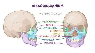
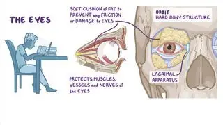
There are two lacrimal bones, each of which is located in the medial wall of the orbit; they are hidden behind the nasal bone when looking at the skull from a frontal view. The orbits, or the eye-sockets, are the two bony cavities that enclose the eyeballs, as well as the muscles, blood vessels, and nerves of the eye. The orbit is pyramidal in shape, with an apex that points towards the inside of the skull, a base that opens out into the facial skeleton, and four walls (i.e., superior, inferior, lateral, and medial) made up of several bones. The medial wall of the orbit consists of the ethmoid bone in the center, the lacrimal and maxillary bones anteriorly, and the sphenoid bone posteriorly. The frontal bone articulates with the lacrimal bone superiorly to form the roof of the orbit, while the lateral wall is made of the zygomatic and sphenoid bones. Finally, the inferior wall, or floor of the orbit, is formed by the maxillary and zygomatic bones, as well as a small part of the palatine bone. 

What is the function of the lacrimal bone?

The main function of the lacrimal bone is to provide structural support to the lacrimal apparatus, or the eye’s tear-producing system. The lacrimal bone is divided by a vertical ridge called the posterior lacrimal crest. This crest creates a hollow depression in the surface of the lacrimal bone (i.e., lacrimal sulcus) that helps form the canal for the nasolacrimal duct. The most superior and dilated part of the nasolacrimal duct is called the lacrimal sac; this is a structure where tears accumulate as they drain from the eye. The posterior lacrimal crest has a hook-like projection called the lacrimal hamulus, which articulates with the frontal process of the maxillary bone, creating an opening for the nasolacrimal duct to drain into the nose. Specifically, lacrimal fluid flows into the inferior nasal meatus, which is located in the inferior nasal concha. This site of drainage explains why individuals typically experience a “runny nose” when they cry. 

In addition, another function of the lacrimal bone is to provide an attachment point for the orbicularis oculi muscle, which is responsible for closing the eyelids and helps promote tear drainage. 

What are the most important facts to know about the lacrimal bone?

The lacrimal bone is a paired facial bone located in the medial wall of the orbit. Its main function is to provide support to the structures of the lacrimal apparatus, which secretes tears to lubricate the eyes, as well as act as a site for orbicularis oculi muscle attachment.
Students say Osmosis is 100% worth it

Because Osmosis saves them time. Lowers stress. And actually helps them remember when it counts.

I used Osmosis to prepare for my first medical school licensing exam! Super helpful and interactive for people who may not do great with just pages of text info!

Cecilia Ruiz

Cecilia Ruiz

MD student

Sayan Misra

I have used Osmosis for about four years. Best thing I have ever used for my medical studies.

Sayan Misra

Sayan Misra

Med student

Osmosis videos are superior because they define simple concepts, tell a story with a clear progression, and provide context.

Jay Pate

Jay Pate

Dental student

References


Drake, R., Vogl, A. W., & Mitchell, A. (2019). Gray’s anatomy for students: With student consult online access (4th ed.). Elsevier - Health Sciences Division.


Hansen, J. T., Netter, F. H. 1., & Machado, C. A. G. (2019). Netter's clinical anatomy (4th ed.). Philadelphia, PA: Elsevier.


Maliborski, A., & Różycki, R. (2014). Diagnostic imaging of the nasolacrimal drainage system. Part I. Radiological anatomy of lacrimal pathways. Physiology of tear secretion and tear outflow. Medical science monitor: international medical journal of experimental and clinical research, 20, 628–638. DOI: 10.12659/MSM.890098