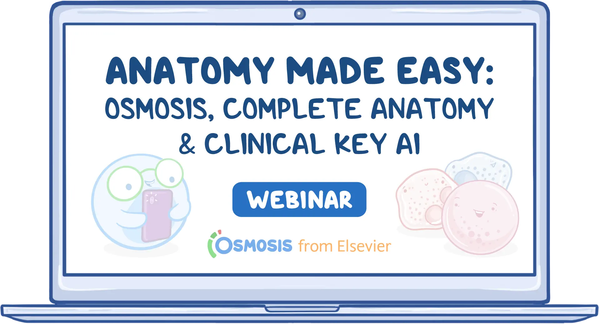
Hi, everyone, and welcome to todays webinar: Anatomy Made Easy: Osmosis, Complete Anatomy, and ClinicalKey AI.
I am Ashton, a medical digital content specialist at Elsevier. My background is in human anatomy, and I have a masters degree from the University of Edinburgh. I joined the Complete Anatomy team and helped create content.
With me today are two Osmosis Healthcare Leadership Initiative students. We have Veronica, a medical surgeon graduate from the Central University of Venezuela in 2023. She is currently on the USMLE path to apply for residency as an internal medicine doctor in the USA. She is part of the Osmosis Healthcare Leadership Initiative and is a regional lead at Osmosis for Elsevier. We also have Asher, a final-year intern medical student at Farabi University in Kazakhstan. He is also an Osmosis Medical Education Fellow. Last year, he completed a clinical elective at Kings College London, and he is aiming to specialize in interventional cardiology.
Here is a quick overview of todays webinar. We are going to look at Osmosis, Complete Anatomy, and ClinicalKey AI. We will dive into each tool with a live demo, explore how each tool supports anatomy learning, and discuss how to use them efficiently. We will also talk about how they can work together, share tips to maximize learning, and touch on additional resources to help you make the most of this webinar.
Why is anatomy such a challenging topic? First, it can feel like information overload. Anatomy requires a lot of memorization, including structures, blood supply, and innervation. It also requires visual and spatial understanding. Understanding these relationships can be complicated when trying to imagine them in a 3D space.
Anatomy also requires integration with clinical knowledge. Anatomy and clinical correlations go hand in hand, and learning these relationships requires both memorization and clinical thinking skills. Retention and application can also be difficult. Students often struggle to retain all the information or understand how it applies to what they will experience clinically.
Terminology is another challenge. When studying anatomy, you are essentially learning a new language. Greek and Latin make up much of the complex language used in healthcare to describe the human body precisely. Lastly, there is simply not enough time. The biggest struggle I hear from students is that they do not have enough time to learn everything. Hopefully, by looking at todays tools, we can help lighten the load.
Today we are going to look at Osmosis, Complete Anatomy, and ClinicalKey AI. Osmosis is video-based and includes clinically integrated content. Complete Anatomy is a 3D anatomy visualization platform. ClinicalKey AI is an AI-powered contextual learning resource.
With that, I will pass it to Asher, who is going to take a deeper dive into Osmosis.
Hi, everyone. I am Asher, and now let us talk about how Osmosis works in action and why students around the world find it so effective.
The first feature of Osmosis is conceptual videos for clear understanding. One of the hardest parts of medical school is taking a complex idea and truly understanding it. Osmosis videos help with that. Instead of overwhelming you with text-heavy slides, they use simple animations, illustrations, and storytelling. This makes even tricky topics easier to follow.
For example, in anatomy, rather than just staring at a list of terms in a textbook, you can see how a nerve or muscle fits into the bigger picture. It feels more like a story than rote memorization, which helps it stick in your mind.
Next are spaced repetition flashcards to boost retention. Anyone who has studied medicine knows that forgetting is part of the struggle. You might study today and feel confident, but two weeks later, the details start slipping away. Osmosis tackles that with smart flashcards. These cards come back to you at the right time, when you are about to forget them. For example, if you learned that the radial nerve controls wrist extension, Osmosis will remind you again and again until it is locked into your long-term memory. This technique has been proven to boost retention, and it saves you from the cycle of cramming and forgetting.
Another important feature is case-based learning for clinical relevance. Osmosis does not show you information in isolation. It connects it to the bigger purpose: caring for patients. Instead of just memorizing the location of a structure, Osmosis shows you why it matters in real life. For instance, if a certain nerve gets injured, how would that affect movement or sensation? These clinical connections keep you engaged and prepare you for the way medicine is applied in reality, not just in exams.
Why do learners love Osmosis? Because it is simple, engaging, and effective. Instead of juggling separate tools for videos, flashcards, practice questions, and case-based learning, Osmosis brings everything together in one platform. It is like having a study partner with you at any time of the day.
Now let us look at a demo of Osmosis and how the features work. On the Osmosis website, you will find a section on the left side for video topics. From there, you can go to Basic Sciences, then Foundational Sciences, and then Anatomy. After clicking on Anatomy, you will find anatomy topics for all regions of the human body.
For this demo, we are looking at the anatomy of the pelvis and perineum. We can go to the anatomy of the pelvic cavity and watch the video. The video includes animations and illustrations and tells the story of the pelvic cavity, including the urinary bladder, rectum, genital organs, and related structures.
There are different sections in the video, so if you want to go to a specific section, you can jump directly to it. You can also turn on captions if needed. Along with the video, you can view the transcript and see the text while you watch. You can also make your own notes alongside the video.
In addition to videos, you can find recall questions to help you actively memorize the topic instead of passively watching. You can start a recall question deck and test your knowledge. For example, one question may ask you to identify the right piriformis by pointing to the image below. You then rate your confidence as low, medium, or high. If you answer incorrectly, the correct structure is highlighted, which helps you learn from the mistake.
You may also be asked to identify the left coccygeus muscle or another structure. Based on your confidence, Osmosis can show you that question again until you remember it. This is how Osmosis helps you recall anatomy.
There are also written questions where you provide answers, rate your confidence, and review explanations. These are very useful for anatomy. Along with the anatomy videos, Osmosis also includes clinically relevant videos. For example, after the anatomy of the pelvic cavity, you can watch clinical correlates related to the female pelvis and perineum. These videos show how anatomy connects to clinical medicine.
The clinical videos also include quizzes and USMLE-style questions. Step 1-style questions focus more on basic sciences, while Step 2-style questions are more clinically focused and involve diagnosis, treatment, and patient management. This means that while you study anatomy, you can also prepare for Step 1, Step 2, or other board-relevant exams.
That is the main overview of Osmosis videos, practice questions, flashcards, and recall questions. You can also explore more features, such as creating your own schedule and custom flashcards. Since we are focusing on anatomy today, I will now hand it over to Veronica to show Complete Anatomy in action.
Thank you so much, Asher. Hi, I am Veronica, and I am going to talk about Complete Anatomy. As Ashton mentioned at the beginning, this is a visualization tool for anatomy and related topics. You can visualize almost everything in detail, and there are also features with videos explaining specific systems or detailed processes, such as the ovarian cycle.
The tools I use the most, especially as I study for Step 1 and the USMLE exams, are the ones that help me observe and retain information. Some key features of Complete Anatomy include custom dissections, the explode tool, layer-by-layer anatomy, cross-sections, saved screens, quizzes and questions, and tools that help you prepare for lab or patient education. It is important not only to know the diagnosis and treatment, but also to understand the pathology a patient is experiencing.
Now we are going to watch a demo of the female reproductive system. From the dashboard, you can go to the bottom left corner and choose the structure or model you want. In this case, we chose the female pelvis and perineum. We can see all the bones of the pelvis, including the ilium, ischium, and pubis. At the bottom, we can see all the systems and add them layer by layer. Here, we are adding muscles, including the levator ani, which is important for the pelvic diaphragm and pelvic support.
I use the app on my tablet because it is easier to move around. Now we have added the urogenital system. Beyond being visually impressive, this complements the Osmosis videos Asher mentioned. Here, we can see the structures and the relationships between them. We can also add the arterial system to see where each artery is located. If we want to add the venous system, we can do that too.
For example, if we look for a specific artery, such as the ovarian artery, we can use a tool on the left that shows origin paths. We have two options: isolate the artery and its origin, which in this case is the abdominal aorta, or highlight the origin path to clearly see the relationship between the ovarian artery and the rest of the system.
If we want to see the female reproductive system specifically instead of layering structures one by one, we can search for it in the top right corner. The app will show the complete female reproductive system. There is a description on the left side where you can read highlights about the system.
One of my favorite tools is the explode tool. This tool helps you see the relationship between structures and even see inside them. It is especially useful for the heart. You can navigate through the whole structure in 3D and understand small structures that can be difficult to distinguish when everything is integrated.
Another useful feature is cross-sections. Complete Anatomy already has cross-sections that you can visualize, but I also like creating personalized cross-sections. In this example, I added the arterial system, nervous system, muscles, and digestive structures. Then I used the cut tool in the upper left corner to create a section through the exact area I wanted to visualize. This lets me see the 3D relationships up close. For example, we can see that the colon is behind the uterus and the bladder is anterior to the uterus.
Since I like this screen, I can save it using the save screen option in the bottom right corner. I can add a screen title so I know what the image is about, save it to My Screens, and tag it under pelvis and perineum. This makes it easy to return to later.
There are also cross-sections already created by the 3D anatomy team. If you go to the library and search for the female reproductive system, you can find pre-made cross-sections. A helpful feature is that these already include labels. You can click on a structure and read its description, which makes studying more interactive and dynamic.
Complete Anatomy also includes questions and quizzes. In the content section of the library, there are questions already made by the team. You can use these to study. If you have something specific you want to retain, you can create your own questions. For example, using the cross-section of the female reproductive system, I can create the question: ?What GI structure is posterior to the uterus?? Then I can add answer choices, mark the correct answer, save the question to My Questions, and tag it under pelvis and perineum.
To create a quiz, I can go to My Questions and drag and drop the questions into a quiz. I can choose options such as shuffling questions or showing correct answers after each attempt. I can also name the quiz so I can find it easily later.
If you pair Complete Anatomy with Osmosis, it is great for moving structures around and visualizing relationships. This is especially important in anatomy lab and in patient education. For example, if you are explaining where an endometrioma might appear, such as in the uterus, fallopian tubes, or pelvic cavity, it is much easier to show it visually than to explain it only with words.
The features I use most are the visualization tool, cut tool, and explode tool. In anatomy, we need to be able to describe, find, and recognize where structures are. Now I will pass it back to Ashton, who will talk about ClinicalKey AI.
Our third tool is ClinicalKey AI, and we are going to look at it in action. Before the demo, some key features are that it is an AI-powered search engine, it has access to trusted anatomy references, and it provides clinical context and explanations based on specific, focused questions. You can also experiment with different phrasing and follow-up questions to refine your search.
When you log in, you will see previous questions on the left if you have used it before. If you want to start fresh, you can select New Conversation. At the top, there is a help button with search tips, a quick product tour, a getting started guide, a support hub, and information about how it works. These resources can help you create better prompts for different types of queries.
ClinicalKey AI should not be used as a clinical calculator or to confirm IV compatibility. You can use natural language and common medical abbreviations in your query, and the platform will display its interpretation of the prompt. It can support basic to complex clinical questions. The more detailed and explicit your question is, the more relevant the response will be.
For example, I can ask, ?What is a stepwise approach to managing postviral cough syndrome?? ClinicalKey AI analyzes the query and retrieves sources. It then provides an AI-generated summary with bullet points. If I want more detail, I can select additional information for a longer text summary.
Throughout the response, you can see references from the ClinicalKey library. If I click details, I can see the section of the source where the information came from. I can also click the reference to go directly to the full text article or source in ClinicalKey. This allows me to verify the information and explore further.
After the references, there is an option to indicate whether the response was helpful. This helps improve future responses. You can also type a follow-up question or select one of the suggested follow-up questions. For example, you could ask about common side effects of montelukast. The tool will analyze the follow-up query, retrieve sources, and provide another referenced response.
It is a straightforward tool, but it can be helpful for clinical situations and for getting quick, source-backed responses. There is also a feedback tab in the top right where you can share suggestions to improve the tool.
Now I will pass it back to Asher.
Now that we know about Complete Anatomy and ClinicalKey AI, we can learn how they work together.
We start with Osmosis. This is where you build your foundation. Think of it as laying the bricks for your medical knowledge. You begin by watching short, high-yield videos that break down concepts into simple, digestible parts. Then you review summary notes to reinforce what you just learned. From there, you test yourself with flashcards and move on to case-based practice questions, such as USMLE-style questions. By the end of that cycle, you have a solid understanding of the topic.
But sometimes, even after learning the concept, you might wonder what a structure really looks like or how it connects to everything else in the body. That is when you move to Complete Anatomy. This tool lets you move beyond 2D diagrams into a fully interactive 3D space. As Veronica showed, you can rotate, zoom, and dissect virtual models to understand how structures relate to one another. It is like having a digital cadaver at your fingertips.
For example, if you have just learned about the brachial plexus in Osmosis, Complete Anatomy lets you visualize the nerves branching out, see their pathways, and understand how an injury in one area might affect the rest. This makes learning clearer and more memorable.
Finally, we use ClinicalKey AI to apply the knowledge in a practical clinical way. Here, you can ask real-world questions, explore clinical cases, and see how anatomy and physiology translate into patient care. For example, you can ask, ?What happens to a patient with a midshaft humeral fracture, and why does it cause wrist drop?? This bridges the gap between theory and practice, turning textbook knowledge into clinical reasoning. You can also review references and learn more about clinical scenarios.
The journey flows in three steps: first, the concept with Osmosis; second, 3D visualization with Complete Anatomy; and third, clinical application with ClinicalKey AI. It is like climbing stairs. Each step builds on the last, and by the time you are done, you do not just know the material; you can see it and apply it in real scenarios.
Why does combining these three tools work better than using them separately?
The first reason is that it covers multiple learning styles. Some people learn best by watching videos, some by interacting with 3D models, and some by solving clinical problems. By combining Osmosis, Complete Anatomy, and ClinicalKey AI, you are not limited to one style. You engage with all of them, which gives the knowledge more ways to stick.
The second reason is that it saves time by reinforcing concepts faster. Normally, if you try to do this on your own, you might watch a lecture, search for 3D references, and then look up clinical cases separately. That takes hours and can leave gaps in understanding. With this integrated approach, you reinforce the same concepts from different angles: video, visualization, and application. This repetition strengthens memory while saving study time.
The third reason is that it builds confidence for exams and patient care. Medical school and exams can feel overwhelming, and many students wonder whether they will remember the material when they need it. By learning in layers, first conceptually, then visually, then clinically, you develop deeper understanding. When you see a patient case or an exam question, you are not guessing. You are recalling knowledge that has been reinforced in different ways. That builds confidence in exams and patient care.
Finally, this approach turns passive learning into active problem solving. Instead of just watching or reading, you are constantly engaging: recalling a flashcard, manipulating a 3D model, or reasoning through a case with ClinicalKey AI. This active process transforms learning from short-term memorization into long-term mastery.
Now I will pass it to Veronica, who will explain active and passive learning and how you can maximize your learning with these tools.
Thanks, Asher. Now we are going to learn about active and passive learning. Osmosis has a helpful video explaining how to study better and how memory works.
When we learn something, we tend to forget it over time. This happens to everyone. Some people say they are slow learners, but forgetting is a normal part of memory. If you learn something on day one, then a week later you may remember only a small portion of what you studied.
The forgetting curve helps us understand why spaced repetition and active learning are important for retaining information. If we learn something on day zero, the curve starts to drop as we begin forgetting. But before we forget completely, we can review the information, use flashcards, or take quizzes. That brings our memory back up. The curve will drop again, but not as sharply as the first time. If we continue reviewing before forgetting, the curve flattens over time, giving us better long-term retention.
That is how spaced repetition and active retrieval work. If we only reread the information, we may recognize it, but we are not truly strengthening retrieval. Actively retrieving it with quizzes and flashcards helps make the information available when we need it, especially for patient care.
To get the most out of these platforms, we need to focus on strategies that promote active learning. Active learning means engaging with the material. We are dissecting models, answering questions, and applying concepts to clinical cases.
Passive learning includes watching videos, reading summaries, or highlighting with different colors. Highlighting may look nice on paper and feel productive, but it does not always help information stick.
For Osmosis, passive learning would be watching videos without taking notes. Active learning would be using spaced repetition flashcards or answering quiz questions. This helps you retain information and identify gaps in understanding. When you answer quiz questions, you can see what you got wrong and what you need to review again.
With Complete Anatomy, passive learning would be watching the 3D models without interacting with them. Active learning means dissecting layers, labeling structures, quizzing yourself, and using the 3D model to explore details. Curiosity helps too. If you move through the structures and explore them because they are interesting, not just because you have to, you are more likely to retain the information.
With ClinicalKey AI, passive learning would be reading summaries without applying them. Active learning means asking clinical questions, comparing differential diagnoses, and using the tool to understand how conditions differ from each other. ClinicalKey AI is useful for this because it can explain specific differences between diseases.
Here are a few additional tips to build better habits. During lectures, it is tempting to write down everything you hear. A more efficient strategy is to write down key concepts, especially high-yield points for exams or patient treatment. Try to listen and understand what the instructor is explaining. Later, when you are studying at home, you can use these tools to revisit anything you did not understand or remember.
Another tip is to study a topic and then quiz yourself right afterward. You can use Osmosis flashcards, Complete Anatomy quizzes you create yourself, pre-made quizzes, or other interactive tools. Complete Anatomy has detailed structures of the eye, ovarian cycle, tongue, and many other regions, and these 3D models help you understand and retain information.
ClinicalKey AI can help with real-life clinical simulations or scenarios, because as healthcare workers, we will treat patients, not just read about them in books. ClinicalKey AI can help connect study topics to real clinical situations.
Short, scheduled sessions are usually better than long, exhausting marathons. I have found that a 30- to 45-minute focused block is more effective. I usually target one region or concept per session, then quiz myself or use flashcards afterward.
Using these tools can help you retain information and retrieve it more efficiently than passive reading. Active learning prepares your brain so that when you need the information, it is available. The final point is consistency. All of these strategies are habits. You can use them and learn a lot, but you need to use them over time for them to make a major difference.
I will now pass it back to Ashton.
Thanks, Veronica. Those were great tips, especially around time management.
Because this is an on-demand webinar, here are a few tips for making the most of it. As you go through the video, pause and follow along with your own accounts, whether that is Osmosis, Complete Anatomy, or ClinicalKey AI. Spend time exploring each tool. You can also go back and rewatch key sections if you need more information.
We also suggest applying what you learn to a specific case or upcoming exam topic while you study. There are additional resources available to help you as well. For Osmosis, there is a help center, a contact page, frequently asked questions, and a blog. For Complete Anatomy, there is a support center, video tutorials to help you learn the platform, and a blog. For ClinicalKey AI, there is customer service support and more information about using the product.
Thank you so much for your interest in this webinar. We hope we have helped you prepare for your upcoming coursework and exams. To access the recording of this webinar or register for future webinars, please visit osmosis.org/events. Thank you again for joining.
Helping current and future clinicians focus, learn, retain, and thrive. Learn more.