Osmosis video - Superficial structures of the neck: Anterior triangle

00:00 / 00:00
More Videos

Anatomy of the larynx and trachea

Anatomy of the lymphatics of the neck
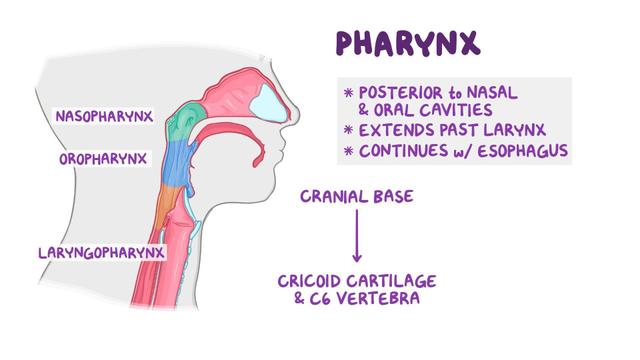
Anatomy of the pharynx and esophagus
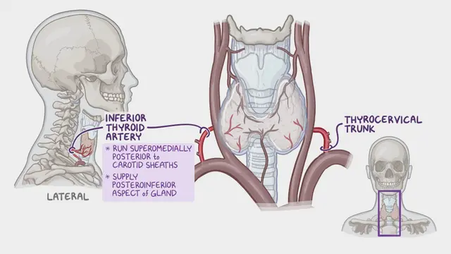
Anatomy of the thyroid and parathyroid glands
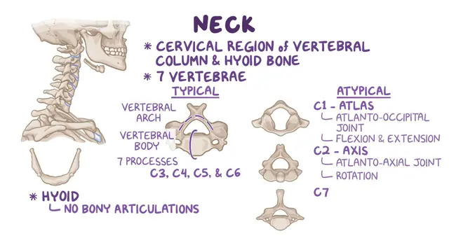
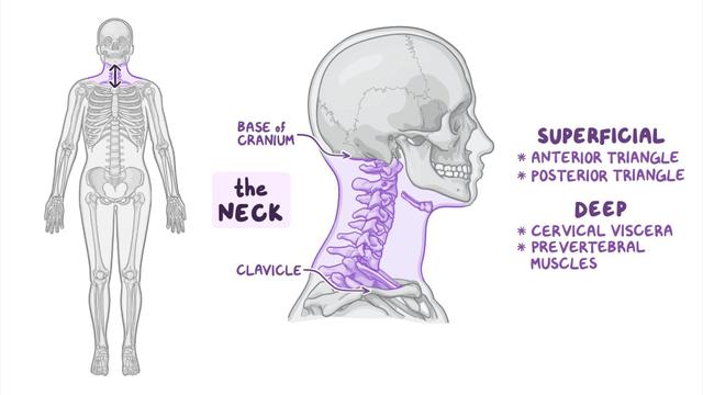
Bones of the neck
Deep structures of the neck: Prevertebral muscles
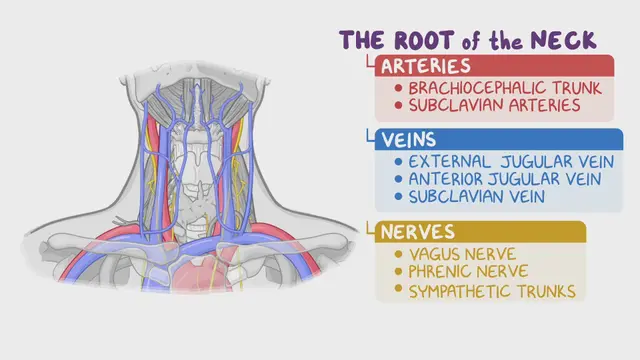
Deep structures of the neck: Root of the neck
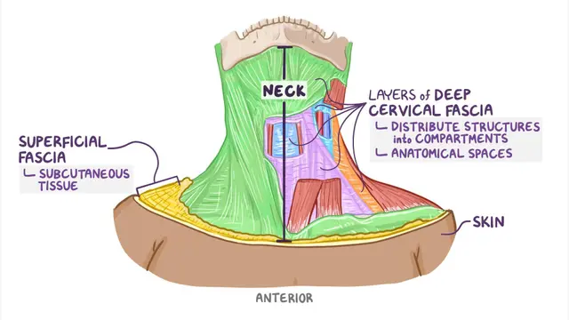
Fascia and spaces of the neck
Superficial structures of the neck: Anterior triangle
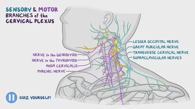
Superficial structures of the neck: Cervical plexus
Superficial structures of the neck: Posterior triangle
Video Summary of Superficial structures of the neck: Anterior triangle
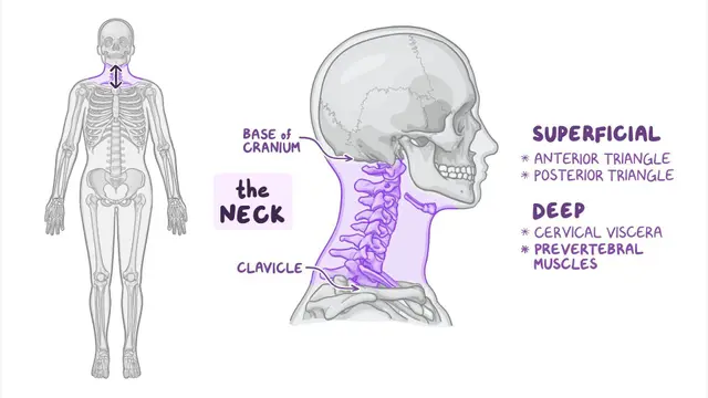
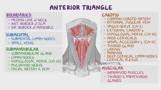
The anterior neck triangle, or just the anterior triangle, is a region of the neck bounded by the inferior border of the mandible superiorly, the anterior border of the sternocleidomastoid laterally, and the sagittal line down the midline of the neck medially.
The anterior triangle is home to several muscles, nerves, arteries, veins, and lymph nodes. Muscles of this region are in two groups: the suprahyoid and infrahyoid muscles. The suprahyoid muscles include the stylohyoid, the digastric, the mylohyoid, and the geniohyoid muscles. The infrahyoid muscles include the omohyoid, the sternohyoid, the thyrohyoid, and the sternothyroid muscles.
Nerves found in the anterior triangle include several cranial nerves, such as CN VII, CN IX, CNX, CN XI, and CN XII. Blood vessels passing through this region include the common carotid artery, which splits into the internal and the external carotid artery supplying various structures in the head. There is also the internal jugular vein, which drains venous blood from the head and neck.