Coagulation Cascade · What Is It, Steps, and More

Published: Sep 10, 2025
Author: Corinne Tarantino, MPH
Editor: Ahaana Singh
Editor: Józia McGowan, DO
Illustrator: Jillian Dunbar
Copyeditor: David G. Walker
7-day free trial

Go deeper with Osmosis

Osmosis is a learning platform with videos, questions, and AI tools to help you master topics like this.

4.8 · 12,000+ reviews
Watch quick, visual videos
Practice with Qbank-style questions
Use AI to explain, quiz, and review
Study anytime with the mobile app
Start free trial

No credit card · Cancel anytime

What is the coagulation cascade?

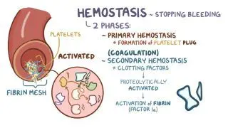
The coagulation cascade, or secondary hemostasis, is a series of steps in response to bleeding caused by tissue injury, where each step activates the next and ultimately produces a blood clot. The term hemostasis is derived from “hem-”, which means “blood”, and “-stasis”, which means “to stop.” Therefore, hemostasis is the process by which bleeding stops. There are two phases of hemostasis. First, primary hemostasis forms an unstable platelet plug at the site of injury. Then, the coagulation cascade (i.e., secondary hemostasis) is activated to stabilize the plug, stop blood flow, and provide time for tissue repair. This process minimizes blood loss after injuries. 

The coagulation cascade involves the activation of a series of clotting factors, the proteins involved in blood clotting. Each clotting factor is a serine protease, an enzyme that speeds up the breakdown of another protein. Clotting factors circulate in their inactive form, known as zymogens. When placed with its co-factor, the clotting factor is activated and is then able to catalyze the next reaction. When a clotting factor becomes activated, it is denoted with an “a” following its respective Roman numeral (e.g., when activated, Factor V becomes Factor Va). 

Learn deeper with Osmosis

Master this topic faster with videos, questions, and AI.

Used by 8M+ healthcare learners.

Start free trial

No credit card · Cancel anytime

What are the three pathways of coagulation?

Coagulation consists of three pathways: extrinsicintrinsic, and common. Together, they interact to form a stable blood clot. The extrinsic and intrinsic coagulation pathways both lead into the final common pathway by independently activating factor X into factor Xa. The extrinsic pathway involves initiation by factor III (i.e., tissue factor) and its interaction with factor VII. On the other hand, factors XII, XI, IX, and VIII are utilized in the intrinsic pathway. Ultimately, the common pathway uses factors X, V, II, I, and XIII to form a stable blood clot. 

The extrinsic pathway is initiated by injury to the endothelial tissue (i.e., skin tissue), exposing and activating tissue factor (i.e., factor III) to the blood. Tissue factor then binds calcium and factor VIIa to activate factor X into factor Xa. Factor VII is present in the blood and requires vitamin K to be activated. 

Meanwhile, the intrinsic pathway begins when factor XII (i.e., Hageman factor) is exposed to endothelial collagen, kallikrein, and high molecular weight kininogen (HMWK) and is subsequently activated into factor XIIa. Factor XIIa activates factor XI into XIa, which in turn binds to a calcium ion and activates factor IX into factor IXa. Then, factor IXa, factor VIIIa, and calcium form a complex to activate factor X to Xa. Factor VIII is found circulating in the blood and is often activated by thrombin (i.e., factor IIa). 

The common pathway is initiated after the activation of factor X into factor Xa at the end of either pathway. The common pathway begins when factor Xa, Va, and calcium bind together, forming a prothrombinase complex. The prothrombinase complex then activates prothrombin (i.e., factor II) into thrombin (i.e., factor IIa), which in turn cleaves fibrinogen (i.e., factor I) into fibrin (i.e., factor Ia). Afterwards, thrombin cleaves the stabilizing factor (i.e., factor XIII) into XIIIa. Factor XIIIa binds calcium to create fibrin crosslinks that stabilize the clot. Thrombin has several functions, including activating platelets (i.e., cell fragments involved in clot formation) and activating factors V, VIII, and IX.  
When is fibrin produced during the coagulation cascade?

Fibrin (i.e., factor Ia) is produced at the end of the coagulation cascade from fibrinogen (i.e., factor I). Fibrin is a long, thin, insoluble protein that forms a mesh-like network to stabilize the blood clot. 

What are coagulation disorders?

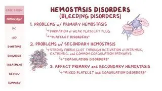
Coagulation disorders are conditions which affect the coagulation cascade, leading to either excessive clotting (i.e., thrombotic disorders) or inadequate clotting (i.e., bleeding disorders). Coagulation disorders usually involve a deficiency in at least one clotting factor, and the most common disorders include von Willebrand disease, hemophilia, and vitamin K deficiency 

Von Willebrand disease is among the most common inherited bleeding disorders and is characterized by a deficiency in von Willebrand factor due to an autosomal dominant genetic mutation. The von Willebrand factor is mostly involved in primary hemostasis, where it helps platelets stick together. The factor also plays a role in secondary hemostasis by helping stabilize factor VIII.   

Hemophilias are bleeding disorders caused by genetic mutations in clotting factor genes. Three types of hemophilia exist: hemophilia A (i.e., factor VIII deficiency)hemophilia B (i.e., factor IX deficiency), and hemophilia C (i.e., factor XI deficiency). All types of hemophilia affect the intrinsic pathway 

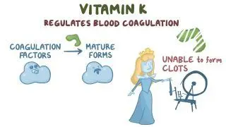
A vitamin K deficiency may occur when an insufficient amount of vitamin K is absorbed from foods, or when not enough foods rich in vitamin K are consumed (e.g., leafy dark green vegetables like spinach). Vitamin K is a cofactor required to activate factors II, VII, IX, and X. Therefore, vitamin K deficiency affects all three pathways. 

What are the most important facts to know about the coagulation cascade?

The coagulation cascade refers to the series of steps that result in the formation of a stable blood clot after injury. It involves a chain reaction of proteins, called clotting factors. Three different pathways (extrinsic, intrinsic, and common) are involved. The intrinsic pathway is activated by factors already circulating in the blood, while the extrinsic pathway is activated by tissue factor. Both pathways result in the activation of factor X to Xa, initiating the common pathway, which ends with the conversion of fibrinogen into fibrin, forming a stable blood clot. Coagulation disorders occur when there is a deficiency in a clotting factor and include von Willebrand Disease, hemophilia, and vitamin K deficiency. 

Key Takeaways

Definition 

A series of steps, also known as secondary hemostasis, which occur in response to bleeding caused by tissue injury, where each step activates the next and ultimately produces a blood clot. It serves to stabilize the platelet plug formed during primary hemostasis. 

The Three Coagulation Pathways 

- Extrinsic pathway 

     - Endothelial/tissue injury exposes tissue factor (factor III) 

     - Tissue factor binds factor VIIa and calcium, forming a complex that activates factor X → Xa 

- Intrinsic pathway  

     - Factor XII → XIIa (contact activation) 

     - XIIa → XI → XIa 

     - XIa + calcium → IX → IXa, which forms a complex with VIIIa + calcium to activate X → Xa 

- Both activate factor X into factor Xa leading to ⤵ 

- Common pathway  

     - Xa + Va + calcium → prothrombinase complex 

     - Prothrombin (factor II) → thrombin (IIa) 

     - Thrombin cleaves fibrinogen (I) → fibrin (Ia) 

     - Fibrin + XIIIa → crosslinked fibrin clot 

Fibrin 

- Produced from fibrinogen at the end of the coagulation cascade 

- Long, thin, insoluble protein  

- Forms mesh-like network to stabilize the blood clot  

Coagulation Disorders 

- Usually caused by deficiency of one or more clotting factor 

- Excessive clotting (thrombotic disorders) 

- Inadequate clotting (bleeding disorders)  

- Von Willebrand disease  

- Hemophilias: 

     - A → factor VIII deficiency  

     - B → factor IX deficiency  

     - C → factor XI deficiency  

- Vitamin K deficiency  

     - Cofactor for factors II, VII, IX, X  

     - Insufficient dietary intake/absorption  

Students say Osmosis is 100% worth it

Because Osmosis saves them time. Lowers stress. And actually helps them remember when it counts.

I used Osmosis to prepare for my first medical school licensing exam! Super helpful and interactive for people who may not do great with just pages of text info!

Cecilia Ruiz

Cecilia Ruiz

MD student

Sayan Misra

I have used Osmosis for about four years. Best thing I have ever used for my medical studies.

Sayan Misra

Sayan Misra

Med student

Osmosis videos are superior because they define simple concepts, tell a story with a clear progression, and provide context.

Jay Pate

Jay Pate

Dental student

References


Kumar, V., Abbas, A., & Aster, J. (2014) Red Blood Cell and Bleeding Disorders. In Robbins & Cotran Pathologic Basis of Disease (9th ed.). Elsevier. 


Ragni MV. Hemorrhagic disorders: coagulation factor deficiencies. In: Goldman L, Schafer AI, eds. Goldman’s Cecil Medicine. 24th ed. W.B. Saunders; 2012:e34–e43. doi:10.1016/B978-1-4377-1604-7.00556-X