
Fusiform Aneurysm · What Is It, Causes, Treatment, and More

Published: Oct 13, 2025
Author: Georgina Tiarks, MD
Editor: Alyssa Haag, MD
Editor: Józia McGowan, DO
Editor: Kelsey LaFayette, DNP, ARNP, FNP-C
Editor: Anna Hernández, MD
Illustrator: Jessica Reynolds, MS
Copyeditor: David G. Walker
7-day free trial

Go deeper with Osmosis

Osmosis is a learning platform with videos, questions, and AI tools to help you master topics like this.

4.8 · 12,000+ reviews
Watch quick, visual videos
Practice with Qbank-style questions
Use AI to explain, quiz, and review
Study anytime with the mobile app
Start free trial

No credit card · Cancel anytime

What is a fusiform aneurysm?

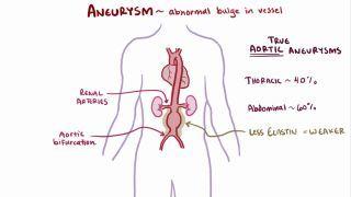
A fusiform aneurysm is a type of aneurysm characterized by a circumferential, balloon-like dilation of a blood vessel. Aneurysms develop when a portion of the blood vessel wall weakens and begins to dilate. To be classified as an aneurysm, the dilation must exceed more than 50% of the vessel’s normal diameter.  

There are many different types of aneurysms based on their shape, including fusiform, saccular or berry aneurysms, and mycotic aneurysms. Fusiform shaped aneurysms balloon outward on all sides and tend to occur in large, high-pressure vessels such as the aorta, whereas a saccular aneurysm only bulges on one side, and typically occur aarterial bifurcations like the Circle of Willis in the brain. On the other hand, a mycotic aneurysm is mushroom-shaped and commonly occurs as a result of an infection, typically bacterial or fungal. 

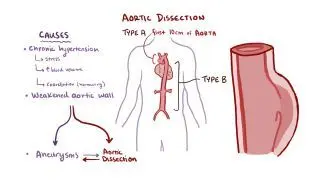
Aneurysms can also be classified based on which layers of the blood vessel wall are involved. Blood vessels are made up of three separate layers: the tunica intima, tunica media, and tunica or adventitia. A true aneurysm is a dilation of all three layers of the vessel wall. In contrast, a pseudoaneurysm, also known as a false aneurysm, occurs when the inner two layers of the vessel wall are disrupted while the tunica adventitia stays intact, allowing blood to pool between the media and adventitia. Finally, a dissecting aneurysm develops when a tear in the tunica intima allows blood to enter the vessel wall, causing the layers to separate. 

Learn deeper with Osmosis

Master this topic faster with videos, questions, and AI.

Used by 8M+ healthcare learners.

Start free trial

No credit card · Cancel anytime

Is a fusiform aneurysm a serious condition?

Yes, fusiform aneurysms can be serious. If they rupture, uncontrolled bleeding may occur, which can be life threatening. 

What causes a fusiform aneurysm?

A fusiform aneurysm is caused by weakening and dilation of the blood vessel walloften resulting from chronic degenerative changes such as atherosclerosis, hypertension, or connective tissue disorders. These conditions result in loss of the elastic fibers and muscle tissue of the tunica media, weakening the structure of the vessel walls. Disorders of elastin and collagen (e.g., Marfan syndrome and Ehlers-Danlos syndrome), inflammatory processes, and certain infections (e.g., syphilis) can also contribute to their formation.  

What are the signs and symptoms of a fusiform aneurysm?

The signs and symptoms of a fusiform aneurysm depend on the location of the aneurysm, size, and whether it is stable, expanding or rupturing. Many fusiform aneurysms, especially those in the aorta, remain asymptomatic for years and are discovered incidentally on imaging performed for other reasons. When symptoms do appear, they usually result from the aneurysm pressing on nearby structures or from compromised blood flow.  For example, an abdominal aortic aneurysm may cause a pulsatile abdominal mass, pain or throbbing in the abdomen or chest, and lower back pain. Similarly, a common iliac aneurysm may result in abdominal or groin pain. A healthcare provider may be able to feel a pulsating mass upon palpation or hear a blowing sound indicating abnormal blood flow (i.e., a bruit) upon auscultation of abdominal, common iliac, or femoral arteries, depending on the location of the aneurysm. 

On the other hand, thoracic aortic aneurysm might present with vague chest or back paina persistent coughhoarseness due to compression of the recurrent laryngeal nerve, or difficulty swallowing if the esophagus is compressed. If the aneurysm affects the aortic valve, it may also result in aortic insufficiency, a condition where the aortic valve doesn’t close completely allowing blood back into the heart with each heartbeat. 

More rarely, fusiform aneurysms may develop in cerebral blood vessels. Unlike saccular aneurysms, which tend to rupture and cause subarachnoid hemorrhagefusiform cerebral aneurysms more often cause symptoms by compressing nearby structures or by reducing blood flow, potentially leading to ischemic stroke. 

The most dangerous complication of any aneurysm is rupture, which constitutes a life-threatening medical emergency due to the potential for massive internal blood loss. When an aneurysm ruptures, it causes sudden, severe pain, often described as tearing or rippingThis is quickly followed by signs of hypovolemic shock, including hypotensiontachycardiacold and clammy skinmental confusion or restlessness, and decreased urinary output due to reduced renal perfusion. 

How is a fusiform aneurysm diagnosed and treated?

A fusiform aneurysm should be attended to immediately as it can become life threatening. For aneurysms located at the abdomen, groin, or extremity, an ultrasound is often used initially to visualize and determine the size of the artery in question. If the aneurysm is large, the individual will often be referred directly for treatment. In other cases, a follow-up computer tomography angiography (i.e., CT angiography) may be used to confirm the diagnosis. Conversely, for a brain aneurysm, conventional angiography may be used to determine the size and location of the aneurysm within the brain. 

Once diagnosed, treatment of an abdominal, groin, or extremity aneurysm will depend on the severity, location, and individual symptoms. With small or asymptomatic aneurysms, conservative treatment may be utilized, which may include ultrasounds to assess for changes, blood pressure control, and management of any acquired risk factors (e.g., smoking cessation, antihypertensive treatment, or medication to lower cholesterol). Conversely, larger or symptomatic aneurysms—or those growing rapidly—typically require surgical or endovascular repair to prevent rupture. Endovascular repair is a less invasive approach in which a stent graft is placed inside the affected blood vessel via catheter to reinforce the weakened wall and exclude the aneurysm from circulation. In cases where surveillance is indicated, regular follow-up visits with a vascular surgeon and scheduled imaging are typically recommended to monitor the size of the aneurysm and progression 

Preventative measures may also be used to detect aneurysms before they enlarge. According to the U.S. Preventive Services Task Force, a one-time screening ultrasound for abdominal aortic aneurysms may be recommended for males aged 65 to 75 years of age who have a history of smoking. Screening for thoracic aortic aneurysms may also be indicated in individuals with connective tissue disorders, a bicuspid aortic valve, or a family history of thoracic aortic aneurysm or dissection. 

What are the most important facts to know about fusiform aneurysms?

A fusiform aneurysm is a circumferential dilation of an artery that affects the entire vessel wall.They most commonly occur in large arteries such as the aorta (especially abdominal or thoracic) and intracranial arteries but can affect any major vessel. Common causes include connective tissue diseases and acquired risk factors such as smoking, hypertension, and atherosclerosis. Diagnosis often involves imaging studies, such as an ultrasound, CT with angiography, or cerebral angiography. Small, asymptomatic aneurysms may be monitored with regular imaging. Larger or symptomatic aneurysms require surgical repair to prevent rupture.  

Key Takeaways

Definition 

A fusiform aneurysm is a type of aneurysm characterized by a circumferential, balloon-like dilation of a blood vessel. 

Seriousness 

- Risk of rupture  

     - Uncontrolled bleeding 

     - Life-threatening 

Causes 

- Weakening and dilation of the blood vessel wall 

- Chronic degenerative changes  

     - Atherosclerosis  

     - Hypertension  

     - Connective tissue disorders  

- Disorders of elastin and collagen  

     - Marfan syndrome  

     - Ehlers-Danlos syndrome  

- Inflammatory processes 

- Certain infections (syphilis) 

Signs and Symptoms 

- Depends on location, size, and if stable, expanding or rupturing 

- Can be asymptomatic for years  

- Symptoms usually start by the aneurysm pressing on nearby structures or from compromised blood flow 

- Abdominal aortic, common iliac, femoral  

     - Pulsating mass upon palpation  

     - Pain or throbbing in the area 

     - Lower back pain

     - Bruit upon auscultation  

- Thoracic aortic 

     - Vague chest or back pain  

     - Persistent cough  

     - Hoarseness 

     - Difficulty swallowing 

- Cerebral (rare)  

     - Deficits from compression of nearby structures 

     - Ischemic stroke from reduced blood flow 

- Rupture  

     - Massive internal blood loss  

     - Sudden, severe pain  

     - Signs of hypovolemic shock (hypotension, tachycardia, cold and clammy skin, mental confusion, restlessness, decreased urinary output) 

Diagnosis and Treatment 

- Immediate care required (life-threatening) 

- Imaging to diagnose 

- Surgical or endovascular repair 

Students say Osmosis is 100% worth it

Because Osmosis saves them time. Lowers stress. And actually helps them remember when it counts.

I used Osmosis to prepare for my first medical school licensing exam! Super helpful and interactive for people who may not do great with just pages of text info!

Cecilia Ruiz

Cecilia Ruiz

MD student

Sayan Misra

I have used Osmosis for about four years. Best thing I have ever used for my medical studies.

Sayan Misra

Sayan Misra

Med student

Osmosis videos are superior because they define simple concepts, tell a story with a clear progression, and provide context.

Jay Pate

Jay Pate

Dental student

References


Cho MJ, Lee MR, Park JG. Aortic aneurysms: current pathogenesis and therapeutic targets. Exp Mol Med. 2023;55(12):2519-2530. doi:10.1038/s12276-023-01130-w 


Golledge J, Thanigaimani S, Powell JT, Tsao PS. Pathogenesis and management of abdominal aortic aneurysm. Eur Heart J. 2023;44(29):2682-2697. doi:10.1093/eurheartj/ehad386 


Isselbacher EM, Preventza O, Hamilton Black J 3rd, et al. 2022 ACC/AHA guideline for the diagnosis and management of Aortic Disease: A report of the American heart association/American college of cardiology joint committee on clinical practice guidelines. Circulation. 2022;146(24):e334-e482. doi:10.1161/CIR.0000000000001106 


Korte J, Marsh LMM, Saalfeld S, Behme D, Aliseda A, Berg P. Fusiform versus saccular intracranial aneurysms-hemodynamic evaluation of the pre-aneurysmal, pathological, and post-interventional state. J Clin Med. 2024;13(2):551. doi:10.3390/jcm13020551 


Screening for abdominal aortic aneurysm: Recommendation statement. Am Fam Physician. 2020;101(10):online-online. Accessed July 29, 2025. https://www.aafp.org/pubs/afp/issues/2020/0515/od1.html