
Open Pneumothorax · What Is It, Diagnosis, and More

Published: Oct 24, 2025
Author: Lily Guo, MD
Editor: Alyssa Haag, MD
Editor: Ian Mannarino, MD, MBA
Editor: Anna Hernández, MD
Editor: Kelsey LaFayette, DNP, ARNP, FNP-C
Illustrator: Jillian Dunbar
Copyeditor: Joy Mapes
7-day free trial

Go deeper with Osmosis

Osmosis is a learning platform with videos, questions, and AI tools to help you master topics like this.

4.8 · 12,000+ reviews
Watch quick, visual videos
Practice with Qbank-style questions
Use AI to explain, quiz, and review
Study anytime with the mobile app
Start free trial

No credit card · Cancel anytime

What is an open pneumothorax?

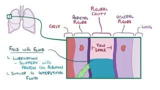
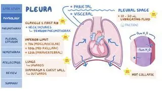
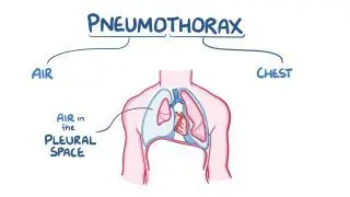
An open pneumothorax happens when air builds up in the pleural cavity, the fluid-filled space between the chest and lungs, due to a hole in the chest wall. When an individual inhales, air enters the pleural cavity through the hole, and this adds pressure on the lung. If the hole is large enough, it may draw enough air into the pleural cavity to make the lung partially collapse, causing shortness of breath 

Learn deeper with Osmosis

Master this topic faster with videos, questions, and AI.

Used by 8M+ healthcare learners.

Start free trial

No credit card · Cancel anytime

What is the difference between an open and closed pneumothorax?

An open pneumothorax and a closed pneumothorax are both types of pneumothorax, the difference between them is how air gets into the pleural cavity 

An open pneumothorax occurs when an injury creates a hole in the chest wall that allows air from the environment to enter the pleural cavity. The most common cause is a penetrating chest injury, such as that from a stab wound or gunshot wound. On the other hand, a closed pneumothorax occurs when air leaks from the lung into the pleural space, but the chest wall remains intact. 

lung disease, such as chronic obstructive pulmonary disease (COPD), cystic fibrosis, lung cancer, or pneumonia, can be the source of a closed pneumothorax. However, damage can also occur spontaneously in predisposed individuals, a phenomenon called spontaneous pneumothorax. 

What causes an open pneumothorax?

An open pneumothorax is often due to forceful impact with a dull object penetrating trauma to the chest wall. Blunt or penetrating trauma can happen in situations such as physical attacks, falls, or motor vehicle accidents.  

In addition, an open pneumothorax may be iatrogenic, meaning it’s a result of a diagnostic procedure or treatment. For example, a clinician could unintentionally injure the chest wall during a transtracheal aspiration, a procedure that requires inserting a needle into the lung tissue; a lung biopsy, in which a portion of the lung tissue is removed; or during a tube thoracostomy, where a tube is placed in the pleural cavity 

What are the signs and symptoms of an open pneumothorax?

The signs and symptoms of an open pneumothorax include sudden chest pain, shortness of breath, rapid and shallow breathing, a fast heart rate, and low oxygenation of body tissues, called hypoxia. Sometimes, individuals with this condition may feel weak, dizzy, restless, or agitated. An open chest wound may be visible, or a sucking sound may be audible when air enters the wound.  

With each breath, air moves freely in and out of the pleural space through the chest wall defect, preventing expansion of the lungs. If air continues to enter the pleural space without being able to escape, the individual may develop a tension pneumothorax. As a result of the rising pressure in the pleural space, the lung collapses and pushes the mediastinum to the opposite side, compressing the heart and the other lung. If left untreated, tension pneumothorax can lead to cardiovascular collapse and cardiac arrest.  

How is an open pneumothorax diagnosed?

An open pneumothorax is usually first diagnosed clinically, especially in trauma settings and then confirmed with imaging. Common examination findings include shallow and rapid breathing and decreased or absent breath sounds on the affected side of the chest.  

Imaging can also assist in diagnosis; the extended focused abdominal sonography for trauma (E-FAST) exam is a diagnostic tool that can be performed rapidly in trauma settings and doesn’t delay initiation of treatment. Computerized tomography (CT) and chest X-ray are also options, although an ultrasound is typically more useful than a chest X-ray as it allows detection of smaller pneumothoraxes. If a patient is unstable with a suspected open pneumothorax, treatment is typically initiated before imaging is used to confirm the diagnosis.  

How is an open pneumothorax treated?

Emergency management of an open pneumothorax involves covering the wound with an occlusive dressing taped on three sides only. This allows air to escape but not re-enter during inspiration, functioning as a one-way valve. If the dressing is not properly applied, and it allows air into the chest space without allowing it to escape, a tension pneumothorax may develop.   

After stabilization, a chest tube may be inserted into the chest wall, typically into the left fifth intercostal space, mid-axillary line. Supplemental oxygen may be required if hypoxia is severe. Long-term treatment involves surgical intervention to repair the open wound or defect in the chest.  

What are the most important facts to know about open pneumothorax?

An open pneumothorax happens when air builds up in the pleural cavity due to a hole in the chest wall. It differs from a closed pneumothorax, during which air enters the pleural cavity from the lungs themselves. An open pneumothorax can occur as a result of direct trauma or unintentional injury in a healthcare setting during a procedure. Symptoms include sudden shortness of breath, pain when breathing, shallow and rapid breathing, and potential shock. Diagnosis involves a thorough examination and confirmation via imaging. Treatment of the pneumothorax often requires application of a three-way dressing and insertion of a chest tube. Surgery may be required to repair the hole in the chest wall that caused the open pneumothorax.  

Key Takeaways

Definition 

An open pneumothorax happens when air builds up in the pleural cavity, the fluid-filled space between the chest and lungs, due to a hole in the chest wall. 

Open versus Closed 

- Open pneumothorax  

     - External injury causes a hole in the chest wall  

     - Air from the environment enters pleural cavity  

- Closed pneumothorax  

     - Chest wall remains intact 

     - Air leaks from the lung into pleural space 

Causes 

- Blunt or penetrating trauma 

     - Physical attacks (e.g. stabbing) 

     - Falls  

     - Motor vehicle collisions 

- Iatrogenic 

     - Due to diagnostic procedure or treatment   

      - Unintentional injury during  

          - Transtracheal aspiration  

          - Lung biopsy  

          - Tube thoracostomy  

Signs and Symptoms 

- Sudden chest pain 

- Shortness of breath 

- Rapid, shallow breathing  

- Tachycardia 

- Hypoxia  

- Weak, dizzy, restless, agitated  

- Sucking sound as air enters wound 

Diagnosis 

- Physical exam 

     - Shallow, rapid breathing  

     - Decreased or absent breath sounds on affected side 

- Imaging 

Treatment 

- Emergency management 

     - Occlusive dressing taped on three sides to serve as one-way valve for air 

- Chest tube 

- Surgical repair 

Students say Osmosis is 100% worth it

Because Osmosis saves them time. Lowers stress. And actually helps them remember when it counts.

I used Osmosis to prepare for my first medical school licensing exam! Super helpful and interactive for people who may not do great with just pages of text info!

Cecilia Ruiz

Cecilia Ruiz

MD student

Sayan Misra

I have used Osmosis for about four years. Best thing I have ever used for my medical studies.

Sayan Misra

Sayan Misra

Med student

Osmosis videos are superior because they define simple concepts, tell a story with a clear progression, and provide context.

Jay Pate

Jay Pate

Dental student

References


Duchman B, Cheng GZ. Management of pneumothorax and persistent air leak—A narrative review. AME Med J. 2024;9(0):23-23. doi:10.21037/amj-23-168 


Fulde S, Fulde GWO. Emergency Medicine: The Principles of Practice. 7th ed. Elsevier; 2020. 


Shorthose M, Barton E, Walker S. The contemporary management of spontaneous pneumothorax in adults. Breathe (Sheff). 2023;19(4):230135. doi:10.1183/20734735.0135-2023 


Tran J, Haussner W, Shah K. Traumatic pneumothorax: A review of current diagnostic practices and evolving management. J Emerg Med. 2021;61(5):517-528. doi:10.1016/j.jemermed.2021.07.006 


Walker S, Hallifax R, Ricciardi S, et al. Joint ERS/EACTS/ESTS clinical practice guidelines on adults with spontaneous pneumothorax. Eur Respir J. 2024;63(5):2300797. doi:10.1183/13993003.00797-2023