Arterial embolism: Nursing
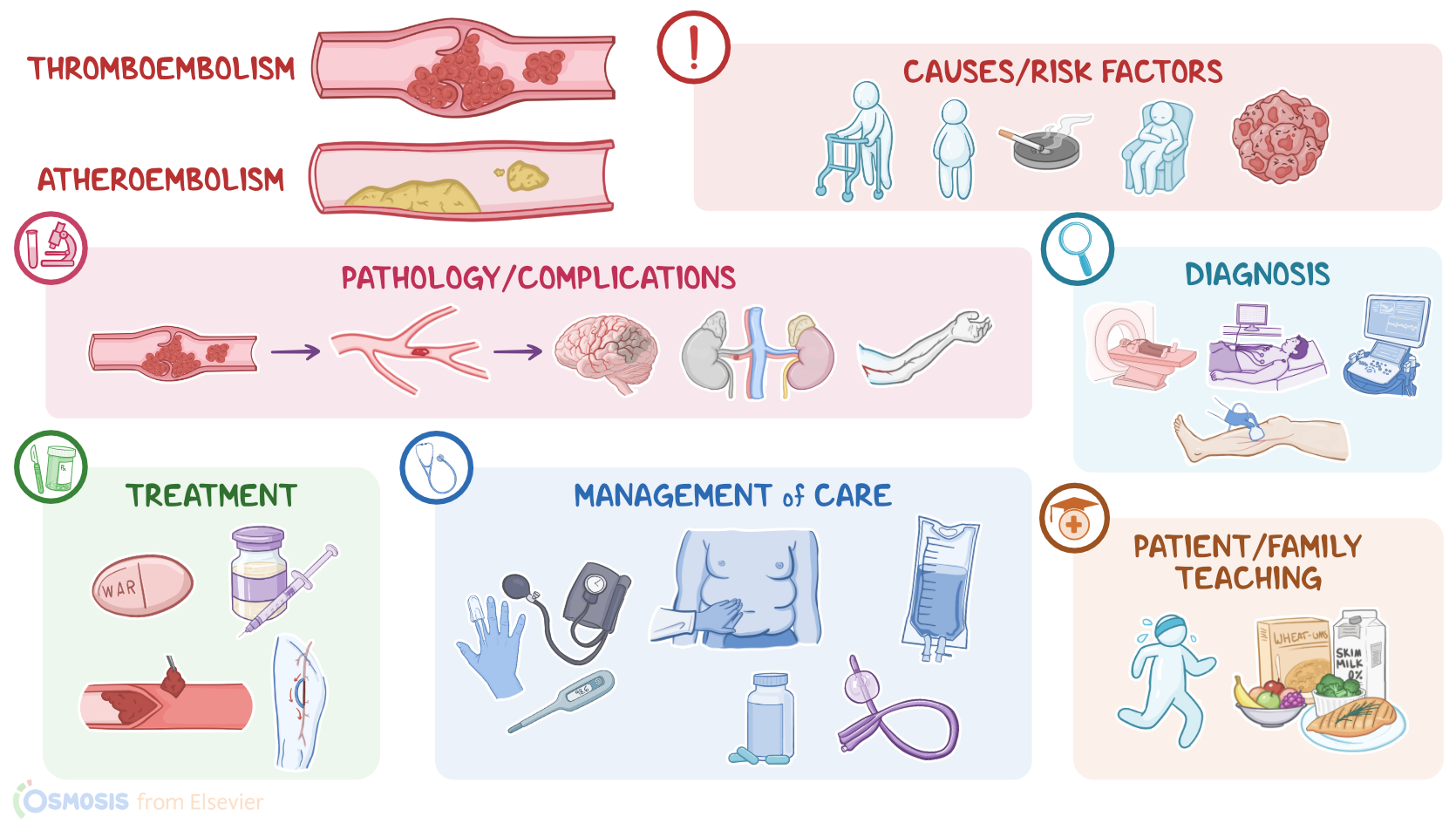
Arterial embolism occurs when some material lodges in an artery, subsequently causing a sudden interruption of arterial blood supply to parts distal to the occlusion. The most common types of arterial embolism include thromboembolism, which occurs when a blood clot, or thrombus, breaks loose, becoming an embolus; and atheroembolism, which occurs when cholesterol crystals from an arterial plaque break loose and enter the circulation.
Alright, let’s cover some basics of physiology. There are three major types of blood vessels: arteries, veins, and capillaries. Normally, blood flows from large arteries into medium and then small arteries called arterioles, which in turn deliver oxygen-rich blood into a network of capillaries, called the capillary bed. Here, oxygen and nutrients pass to tissue cells, which will then return carbon dioxide and wastes back into the capillary bed. These capillaries will then merge to form the smallest veins, called the venules. Eventually, these venules converge to form small veins, which then drain into medium superficial veins. Next, these superficial veins drain blood into medium deep veins, which continue to converge, forming large veins that eventually drain back into the right side of the heart. From here, venous blood goes into the lungs, where it gets oxygenated.
Now, let's zoom in and take a closer look at the arterial wall, which consists of three layers: from outside in, there’s tunica externa or the adventitia layer, which has loose connective tissue; then tunica media or the middle layer, which contains some elastic tissue and smooth muscle that allow the artery to dilate or constrict in response to local conditions; and finally the endothelium, which consists of a single layer of endothelial cells on top of a layer of connective tissue, called lamina propria. The endothelial cells maintain blood flow by preventing blood cells from reaching the underlying lamina propria. Additionally, endothelial cells express small molecules such as heparin sulfate and prostacyclin, which help prevent blood coagulation. So, if their continuity is disrupted, blood cells may come up against coagulation factors in the lamina propria. This initiates a coagulation cascade that leads to the formation of a blood clot, which in turn blocks the blood flow.
| ARTERIAL EMBOLISM | ||
| KEY POINTS | NOTES | |
| DEFINITION |
| |
| PHYSIOLOGY |
| |
| CAUSES AND RISK FACTORS |
| |
| PATHOPHYSIOLOGY |
| |
| SIGNS AND SYMPTOMS |
| |
| DIAGNOSIS |
| |
| TREATMENT |
| |
| MANAGEMENT OF CARE |
| |
| PATIENT AND FAMILY TEACHING |
| |
No notes for this video yet
Try adding a note below