Bell palsy: Nursing
Introduction0:00–0:14
Bell palsy is a condition characterized by weakness or paralysis of the muscles on one side of the face, that’s caused by damage to cranial nerve VII, which is the facial nerve.
Now, let’s quickly review the anatomy and physiology of facial innervation. The 12 pairs of cranial nerves are peripheral nerves, also called lower motor neurons, that emerge from the brain or brainstem.
Physiology0:14–2:06
The seventh cranial nerve is the facial nerve, which emerges from the brainstem, and then enters the temporal bone where it travels through the facial canal.
The facial nerve then exits the skull through a tiny hole called the stylomastoid foramen. From there, it branches off to innervate different facial muscles that control facial expression ipsilaterally, so on the same side.
The facial nerve also innervates the lacrimal glands, as well as the sublingual gland and submandibular salivary glands, and the mucous membranes of the nose, mouth, and nasopharynx.
In addition, the facial nerve also carries sensory information about taste from the anterior ⅔ of the tongue. Finally, the facial nerve innervates the stapedius muscle in the ear, which helps dampen loud noises.
If we look back, the facial nerve receives information from a region of the brain called the motor cortex, which has upper motor neurons that send information to the brainstem nuclei of the facial nerve, and then the facial nerve gives rise to two lower motor neurons, one to innervate the lower side of the face, and one for the upper side.
Now, the lower motor neuron that innervates the upper side of the face receives information from both sides of the motor cortex, so both from an upper motor neuron coming from the ipsilateral side, as well as an upper motor neuron coming from the contralateral or opposite side that crosses in the midline.
On the other hand, the lower motor neuron that innervates the lower side of the face only receives information from the contralateral motor cortex, so from an upper motor neuron that crosses in the midline.Now, Bell palsy is caused by damage to the facial nerve.
Causes & risk factors2:06–2:42
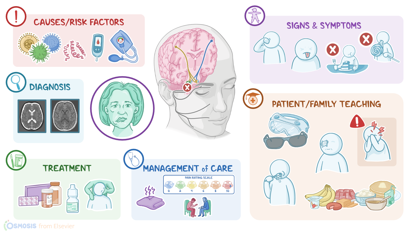
Although the precise cause of the damage is unknown, risk factors include history of viral infections, such as herpes simplex virus or HSV, Epstein-Barr virus or EBV, and varicella-zoster virus or VZV; as well as bacterial infections, such as Borrelia burgdorferi, which causes Lyme disease.
Other risk factors include conditions like sarcoidosis, diabetes mellitus, or hypertension.So, these risk factors seem to trigger inflammation or damage to the lower motor neurons belonging to the facial nerve.
Pathology2:42–3:11
As a result, information from both the contralateral and the ipsilateral motor cortex is lost for the upper face, as well as information from the contralateral motor cortex for the lower face.
This results in the paralysis of all the muscles on the ipsilateral side of the affected nerve.Now, the clinical manifestations of Bell palsy typically begin suddenly, and tend to worsen over 48 hours.
Clinical manifestations3:11–4:26
These include mild weakness to total facial paralysis on one side; as well as a unilateral drooping of the eyelid and mouth, and disappearance of the nasolabial fold, which is the skin fold that runs from the side of the nose to the corner of the mouth.
Clients often experience dryness of the affected eye or side of the mouth, a loss of taste sensation on the anterior ⅔ of the tongue, as well as some difficulty drinking and eating, or difficulty with speech.
In addition, clients often experience posterior auricular pain, or pain behind the ear. Lastly, some clients can also develop unilateral hyperacusis, where the ear on the affected side may have increased sensitivity to loud noises.Now, it’s important to distinguish Bell palsy from other causes of facial paralysis, where the underlying problem is in the motor cortex, before the upper motor neurons cross the midline.
This only causes paralysis of the lower half of the face on the contralateral side of the lesion, since the upper half of the face is still receiving some information from the ipsilateral motor cortex.The diagnosis of Bell palsy starts with the client’s history and physical assessment, followed by brain imaging tests, such as MRI or CT scan, in order to rule out a stroke or brain tumor.Regarding treatment for Bell palsy, most cases resolve on their own within 6 months, but some clients may benefit from anti-inflammatory medications, like corticosteroids, which can help reduce the nerve inflammation and speed up the recovery.
Diagnosis4:26–4:43
Treatment4:43–5:11
In some cases, physiotherapy can be used after recovery to promote facial muscle control. Fortunately, only a few clients develop permanent facial weakness or paralysis.Okay, let’s look at the nursing care you’ll be providing for a client with Bell palsy.
Management and care5:11–6:15
The priority goals of care include managing symptoms and preventing complications.Begin by assessing if your client is experiencing pain, and administer the prescribed analgesics and apply a warm, moist compress to the affected side to provide some additional comfort and promote circulation.
Then, administer the prescribed corticosteroids. Next, assess their eyes and administer artificial tears or ointment to keep the cornea from drying out.
Lastly, collaborate with the physical or occupational therapist who may recommend and fit a facial sling for your client to help provide support, improve lip alignment, and help with eating.Finally, provide support for your client regarding their temporary change in physical appearance, and encourage them to express their feelings and anxieties.
Refer them to counseling, as needed. Alright, let’s move on to client and family teaching.
General client and family teaching6:15–8:27
Begin by explaining how the disorder affects their facial nerve, resulting in the symptoms they are experiencing. Reassure them that in most cases, the condition is self-limiting, and that their symptoms should resolve gradually over several months.
Lastly, emphasize the importance of keeping all scheduled treatment and follow-up appointments so their progress can be monitored.
Next, discuss the importance of protecting their eyes. Instruct them on the proper use of the moisturizing eye medications during the day, and explain that wearing protective eyewear like goggles or sunglasses will help protect their eye from dirt and dust, since they may not be able to blink to protect their eye.
Also let them know that they can close their affected eye manually several times each day to simulate blinking. Demonstrate how to apply ointment at night, to gently tape the affected eyelid shut, and then apply a patch at night to protect their eye during sleep.
Instruct them to contact their healthcare provider right away if they experience any symptoms of corneal abrasion, including itching, pain, irritation, and light sensitivity, vision loss, or a feeling like there’s something gritty in their eyes.
Stress the importance of keeping their follow-up appointments with their ophthalmologist to monitor their eye health.Also be sure to teach your client about strategies for managing pain and discomfort.
Recommend they apply moist heat to the affected area to promote circulation as needed, and take their prescribed analgesics and corticosteroids as directed.
Also review changes they’ll need to make while eating. Teach them to chew on the non-affected side of their mouth to prevent food trapping and advise them to choose soft foods that are easy to chew.
Emphasize the importance of maintaining good oral hygiene after every meal to prevent cavities, periodontal disease, and parotitis.
Remind them to maintain their scheduled dental cleanings every 6 months to monitor for any problems with their oral health.
Finally, as they recover, encourage them to seek support from friends, family, or from a support group.Alright, as a quick recap ….
Review8:27–9:44
Bell palsy is a condition where temporary weakness or paralysis of the muscles on one side of the face occurs because of damage to cranial nerve VII, the facial nerve.
Risk factors include a history of viral or bacterial infections or conditions like diabetes mellitus and hypertension that can trigger inflammation or damage to the lower motor neurons of the facial nerve.
Signs and symptoms usually occur suddenly and worsen over 48 hours, and include weakness or facial paralysis on one side, and unilateral drooping of the eyelid and mouth, and disappearance of the nasolabial fold.
Clients often also have dryness of the eye or mouth on the affected side, difficulty talking and eating, and loss of taste.
Diagnosis involves history and physical assessment followed by brain imaging to rule out a stroke or brain tumor. Treatment includes corticosteroid medications to reduce nerve inflammation, as well as physiotherapy after recovery to promote facial muscle control.
Priority goals of care include managing symptoms and preventing complications. Client and family teaching focuses on management of symptoms
| BELL PALSY | ||
| KEY POINTS | NOTES | |
| DEFINITION |
| |
| PHYSIOLOGY |
| |
| CAUSES AND RISK FACTORS |
| |
| PATHOPHYSIOLOGY |
| |
| SIGNS AND SYMPTOMS |
| |
| DIAGNOSIS |
| |
| TREATMENT |
| |
| MANAGEMENT OF CARE |
| |
| PATIENT AND FAMILY TEACHING |
| |
No notes for this video yet
Try adding a note below