Brain tumors: Nursing
Notes
| BRAIN TUMORS | ||
| KEY POINTS | NOTES | |
| DEFINITION |
| |
| PHYSIOLOGY |
| |
| CAUSES AND RISK FACTORS |
| |
| PATHOPHYSIOLOGY |
| |
| SIGNS AND SYMPTOMS |
| |
| DIAGNOSIS |
| |
| TREATMENT |
| |
| MANAGEMENT OF CARE |
| |
| PATIENT AND FAMILY TEACHING |
| |
Transcript
Brain tumors are abnormal growths that can be broadly classified into primary tumors, which originate from cells within the nervous system, and secondary or metastatic tumors originating and spreading from cells outside the nervous system.
Okay, let’s start by looking at the physiology of the nervous system, which is divided into the central nervous system, that includes the brain and spinal cord, while the peripheral nervous system includes all the nerves that connect the central nervous system to the muscles and organs.
Zooming in, the brain is made up of different types of cells. First, there are neurons, which receive and send electrical impulses to one another. Then there are neuroglial cells, which help support and protect the brain, and can be classified into astrocytes, which are the most abundant, as well as ependymal cells, microglia, and oligodendrocytes.
Astrocytes have long processes with enlarged terminal ends that surround blood vessels of the brain as part of the blood-brain barrier. so that only certain molecules can slip through. Astrocytes also help provide nourishment to neurons, and recycle neurotransmitters. Next are ependymal cells, which line up the ventricles of the brain, and produce CSF. Then, microglia are the immune cells of the central nervous system. Lastly, there are oligodendrocytes, which have cellular processes that wrap themselves around axons to form a myelin sheath. In the peripheral nervous system, the myelin sheath is similarly formed by another type of cells, called Schwann cells. Additionally, there are cells that secrete hormones into circulation. These cells are found in the pituitary gland, located at the base of the brain.
Some brain cells have a limited ability to be replaced, especially after injury, and they do it by having undifferentiated stem cells, called neural stem cells and glioblasts, which activate and mature into neurons or neuroglia, respectively.
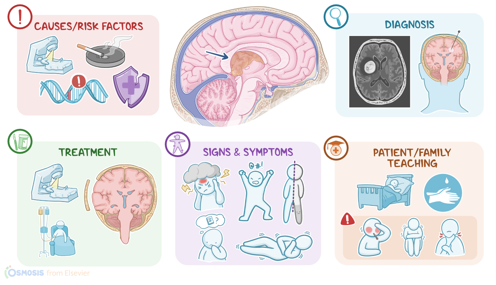
Now, brain tumors occur when any of these cells acquire mutations, which can arise due to a variety of risk factors. Environmental risk factors include exposure to ionizing radiation, which can come from radiotherapy for cancer, or diagnostic imaging like CT scan or MRI. Another risk factor is exposure to N-nitroso compounds found in tobacco, beer, and food products treated with nitrite like fish or cured meats. Genetic risk factors include hereditary conditions like tuberous sclerosis, neurofibromatosis, and von-Hippel-Lindau disease, where affected clients are genetically predisposed to develop brain tumors even without the presence of environmental risk factors. Finally, immunocompromised clients, like organ transplant recipients or clients with AIDS are at higher risk for certain types of brain tumors.
Okay, so the pathology of brain tumors begins once a cell of the brain becomes mutated and starts dividing uncontrollably, forming a tumor. As the tumor grows in size, it can compress nearby structures, interrupting their normal functions, and leading to focal neurological deficits. A tumor found near the ventricles can also block the flow of cerebrospinal fluid, causing hydrocephalus. Now, a rapidly growing tumor will need to get supplied by new blood vessels that are more fragile and can rupture, causing acute bleeding. In addition, these blood vessels are typically not regulated by the blood-brain barrier, which may result in brain edema. And since the cranial cavity is a hard, non-expanding shell, the combined mass effect of large tumors, hydrocephalus, and brain edema, can lead to an increased intracranial pressure. If left untreated, this can result in herniation of the brain and even death.
Now, brain tumors are classified based on the type of cell from which they arise. In adults, the most common ones are gliomas, which are further divided into astrocytomas, such as glioblastoma multiforme, and oligodendroglioma; there’s also schwannomas that most often arise from the Schwann cells ensheathing the acoustic nerve; meningiomas that arise from the meninges; and hemangioblastomas arising from blood vessels.
Another type of brain tumor is the pituitary adenoma, which can be functional, meaning it secretes certain hormones, causing hyperpituitarism; or it can be nonfunctional.
In this case, the tumor might grow to a size that ends up compressing nearby structures, as well as the rest of the pituitary gland, preventing it from functioning correctly, and thus causing hypopituitarism.
Finally, primary central nervous system lymphoma is another type of brain tumor, which is most common in immunocompromised clients, and is caused by the Epstein-Barr virus, or EBV. On the other hand, gliomas are the most common ones in children, like pilocytic astrocytomas, and ependymomas; and there’s also medulloblastomas, which arise from stem cells; and craniopharyngioma, which originates from embryonic remnants.
Now, the clinical manifestations of brain tumors vary based on the type, size, and location. Common symptoms include a severe headache that may awaken the client at night, as well as seizures, and cognitive dysfunction that may lead to memory and personality changes. Focal neurological deficits can also develop, leading to symptoms like hemiplegia, aphasia, and visual impairment.
In cases of increased cranial pressure, clients can experience nausea and vomiting, or present with late signs like hypertension, bradycardia, and irregular breathing; these signs are referred to as a Cushing reflex.
Finally, clients with functioning pituitary adenomas may present with symptoms that result from excess pituitary hormone levels, such as acromegaly or gigantism, infertility, or Cushing disease.
Diagnosis of brain tumors begins with the client’s history and physical assessment, followed by imaging tests, like an MRI or a contrast CT scan to visualize the tumor. Once a suspicious lesion is found on imaging, a biopsy is needed to confirm the diagnosis, as well as to assess the aggressiveness and prognosis.
So instead of the TNM system, brain tumors are graded according to the WHO classification, which takes into account how tumor cells appear under the microscope. The higher the grade, the more aggressive the tumor is. Grading is a four-stage system, G1, G2, G3, and G4 that evaluates the cell differentiation, meaning how different the cancer cells look compared to the healthy tissue cells. G1 is the most differentiated, meaning that the cancer cells look similar to healthy cells, and G4 is the most undifferentiated, meaning that the cancer cells look totally different than healthy cells. Each type of tumor can have a different grade, but in general, some tend to be low grade, like oligodendrogliomas, schwannomas, meningiomas, and hemangioblastomas; whilst others are typically high grade, like glioblastoma or medulloblastoma.