Cardiac biomarkers - Troponin: Nursing
Cardiac biomarkers - Troponin: Nursing
Acute Final
Acute Final
Notes
| CARDIAC BIOMARKERS - TROPONIN | ||
| KEY POINTS | NOTES | |
| PHYSIOLOGY |
| |
| PATHOLOGY |
| |
| INDICATIONS |
| |
| NURSING IMPLICATIONS |
| |
Transcript
A 50-year-old client is brought to the emergency department by ambulance complaining of chest pain at rest and shortness of breath for the past 30 minutes. His medical history is significant for smoking and hypertension. An ECG reveals new T-wave inversions. The health care provider suspects acute coronary syndrome and orders a troponin level to be drawn.
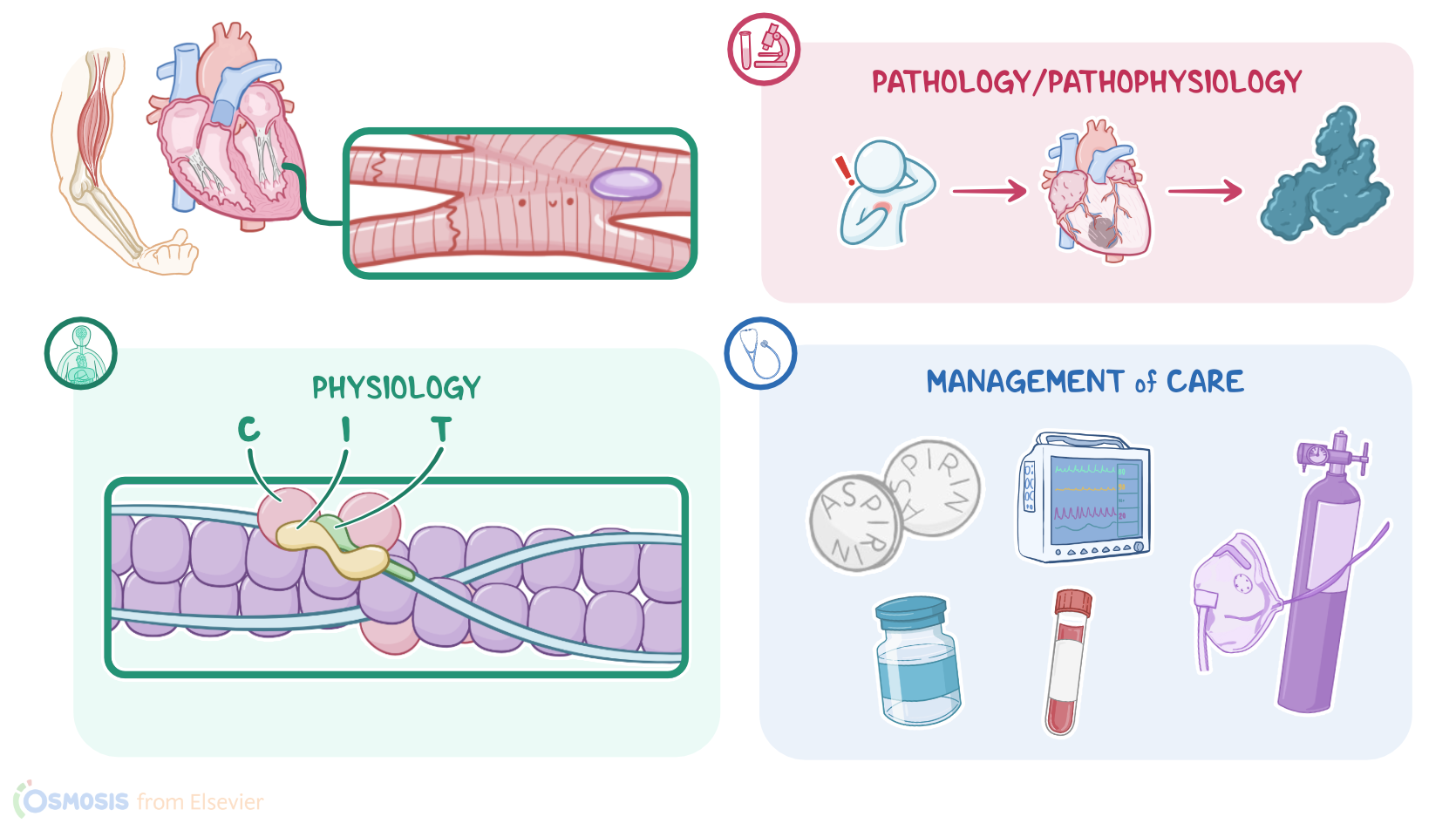
Alright, troponin is a protein found in striated muscles, including skeletal muscles and the myocardium of the heart. There are three types: C, I, and T. Troponins I and T, are highly specific to myocardial tissue, and together, they are known as cardiac troponins, or cTn for short.
Now, cardiac muscle cell, or myocyte, damage can occur with acute coronary syndrome, which is any condition that decreases blood flow to the heart muscle, such as angina or a myocardial infarction, or MI for short. Decreased blood flow causes an imbalance between myocardial oxygen demand and supply from the coronary arteries, resulting in myocardial ischemia and damage to cardiac muscle cells. As the cell membranes become damaged, cellular contents are released into the bloodstream, including proteins like cTn, and enzymes, like creatine kinase-MB, or CK-MB for short, which is found mostly in heart muscle cells.
Alright, so troponin levels will be measured when there is a concern about cardiac ischemia.
Troponin levels are usually drawn together with other cardiac biomarkers like CK-MB. Typically, a blood sample is drawn on admission and repeated to monitor for trends in the values, or if there are new signs and symptoms of ischemia, like new findings on ECG or evidence of new abnormalities on imaging tests.
High-sensitivity cTn assays, or hs-cTn for short, can detect very small levels of cardiac troponin, so cardiac injury can be rapidly identified or ruled out. Individual troponins can also be drawn; the normal range for troponin I is 0 to 0.04 ng/mL, and for troponin T it is 0 to 0.01 ng/mL. Troponin levels can be elevated in the blood within two to four hours after the onset of cardiac ischemia, and usually peak at around 48 hours. After that, they remain elevated for about 7 to 10 days.