Fractures: Nursing process (ADPIE)
Client Report0:00–0:34
Malaya Tanglao is a 15 year old female who presented to the ED with her father Ulan after being unable to bear weight on her left lower extremity following a skiing accident.
X-ray confirms that she sustained a closed transverse mid-shaft fracture to her left tibia that is displaced with resulting soft tissue injury.
Malaya is now being admitted to the pediatric orthopedic unit for pain management and preparation for surgical intervention.
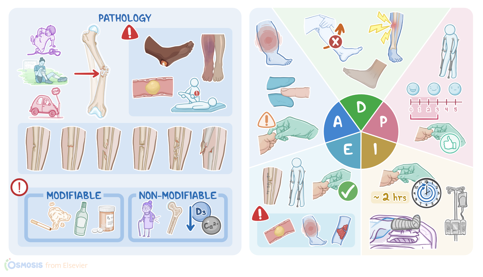
A fracture is defined as a complete or partial break in a bone, which occurs when the physical force applied to the bone is stronger than the bone itself.
Pathology0:34–6:23
Most often this occurs due to trauma associated with a fall, sports injury, or motor vehicle crash. Fractures may also result from overuse during repetitive activities such as running or jumping.
Finally, some conditions such as osteoporosis or cancer can weaken the bones and cause spontaneous fractures. There are some factors that can put an individual at risk for fractures.
Modifiable risk factors that may weaken the bones include low Vitamin D smoking, alcohol, and glucocorticoid use, while non-modifiable risk factors include increasing age, as well as congenital disorders like osteogenesis imperfecta and malabsorption problems that may impair the ability to absorb important nutrients for bone health, like calcium and Vitamin D Now, most commonly we talk about closed or simple fractures, which occur when the bone breaks, but the overlying skin remains intact.
On the other hand, open or compound fractures occur when the fractured ends pierce through the overlying skin. There are many different types of fractures, such as green stick fractures, which occur when one side of the bone breaks while the other side of the bone bends.
Impacted fractures occur when a piece of one bone gets wedged into another bone. Commminuted fractures are where the bone breaks into multiple fragments.
Finally, in spiral fractures, the fracture line follows the projection of a strong twisting force that is applied to the bone.
Spiral fractures are most commonly seen in non-accidental traumas such as physical abuse, like when someone forcefully grabs and twists an arm.
Now the broken bone typically requires several weeks to months to heal. The process starts with the inflammatory phase in which the body responds to the trauma by sending immune cells to the location of the fracture.
Immune cells remove dead and damaged tissue, thereby enabling the body to enter the second phase called the reparative phase.
In this stage, the body activates osteoblasts to form a callus, which is a new bone tissue that connects the fractured ends.
In the last phase, which is also known as the remodeling phase, the callus is replaced by regular bone and the healing process is completed.
Now, fractures typically present with signs and symptoms like localized pain, especially when trying to move, as well as swelling and bruising.
If the fracture is displaced, the affected part of the body can look misaligned, shortened, or even deformed. Fractures can also lead to some serious complications.
Soon after the injury, the broken ends of the bone may damage surrounding structures such as blood vessels, resulting in bleeding, as well as nearby nerves leading to altered sensation and tears to the muscles or tendons.
Another acute complication is compartment syndrome, which is when bleeding or edema from the fracture leads to increased pressure inside the section of the limb that contains muscles, nerves, and blood vessels and results in the reduction of blood supply and tissue necrosis.
Fractures of long bones may lead to fat embolism, which is when a piece of fat breaks off from the fractured bone and then travels through the bloodstream and gets lodged within a blood vessel in organs like the heart, lungs, or brain, obstructing their blood flow.
Fractures may also have long-term complications such as healing abnormalities that may result in bone deformity. This includes malunion, which occurs when the fractured ends are not adequately aligned, delayed union, which is when a bone requires more time to complete the healing process, and non-union, which occurs when a bone completely fails to connect broken ends of the bone.
Some clients might also develop mobility complications such as joint stiffness or instability. On rare occasions, they can also develop contractures where muscles and tendons get shortened and the range of motion gets limited.
Finally, clients who are immobilized and are spending too much time in bed can develop a pressure injury which typically occurs over bony prominences like the sacrum or heels.
Immobilized individuals are also at risk for developing deep vein thrombosis, or DVT for short, which typically occurs in the deep veins of the lower extremities.
The blood clot can break off and travel all the way to the lungs, ultimately causing a pulmonary embolism. Diagnosis of fractures is typically done performing an X-ray in two different planes, most commonly anteroposterior and lateral planes.
If the X-ray finding appears normal and the clinical presentation suggests a possible fracture, MRI or CT should be used to confirm the diagnosis.
Treatment of bone fractures generally involves rest and immobilization of the affected limb in addition to the use of removable splints or casts to allow the bone to heal.
Closed reduction refers to the alignment without surgical intervention, while open reduction is done with surgery. Once aligned, bones can be held in place using internal and external fixation.
On the other hand, in external fixation, pins, screws, and wires are inserted into the fractured bone and held in place by a bar or ring outside of the skin.
Assessment6:23–8:04
Malaya and Ulan have arrived at the pediatric orthopedic unit. After introducing yourself, confirming Malaya's identity, and performing hand hygiene, you begin your assessment by noting Malaya's stiff body posture and how she holds her breath with any physical movement.
Her vital signs are temperature 98.2 °F or 36.7 °C, heart rate 94 BPM, R 16 breaths per minute, and BP 119/62 millimeters of mercury, and SPO 2 94% on room air.
Maleaa rates her pain 6 out of 10 and tells you it doesn't hurt too bad when I stay still, but if I move, I get a sharp shooting pain up and down my left leg.
Her capillary refill is less than 3 seconds. Pedal pulse is strong and palpable, and she is able to wiggle her toes.
Malaya is also able to feel your touch and denies experiencing any numbness to the affected area. At this time, Ulan expresses concern about Malaya's limited mobility to her left lower extremity and how she'll be able to safely get around at school.
After documenting your assessment, you let Malaya and Ulan know the orthopedic surgeon will be in soon to obtain surgical consent.
Based on the data you collected through your assessment, you create these priority nursing diagnoses acute pain related to left tibia fracture, risk for peripheral neurovascular dysfunction related to disrupted bone and soft tissue integrity, and impaired physical mobility related to left tibia fracture.
Diagnosis8:04–8:24
Planning8:24–8:51
It is now time to create a plan of care with the multidisciplinary healthcare team that will be caring for Malaya throughout her hospitalization.
The following goals are established. During her hospital stay, Malaya's pain level will be managed at her stated tolerable level of 3 out of 10 or less, and she will continue to have adequate circulation and full sensation to her left lower extremity.
Before she is discharged, she will be able to safely mobilize on crutches. Next, you begin implementing the plan of care.
Implementation8:51–9:43
You initiate pain management by administering the ordered IV analgesic and teach Malaya and Ulo about non-pharmacological ways to relieve pain, such as ice packs, pursed lip breathing, and listening to music.
Next, you ensure that Malaya's left lower extremity is properly immobilized with the current splint and elevated on pillows to increase peripheral circulation and decrease edema.
You perform a CMS check every 2 hours to monitor for signs and symptoms of neurovascular compromise. While assisting Malaya to ambulate to the bathroom, you teach her the proper use of crutches, as well as the importance of being non-weight bearing on her left lower extremity.
You also inform Malaya and Ulan that a physical therapist will work with Malaya postoperatively before she discharges. Malaya had a successful open reduction and internal fixation of her left tibia and has been transferred back to the pediatric orthopedic unit for continued monitoring and evaluation.
Evaluation9:43–11:02
Her vital signs are temperature 98.5 °F or 36.9 °C, heart rate 70 BPM, respirations 14 breaths per minute, BP 110/66 millimeters of mercury, and oxygen saturation 98% on room air.
Malaya is rating her pain 2 out of 10 and is resting comfortably with her left lower extremity elevated on pillows. Malaya's CMS remains intact, and she tells you that her new cast is not feeling too tight.
The physical therapist has checked in with you and plans to evaluate her in a few hours. Ulan reports that he has contacted Malaya's school and arranged the necessary accommodations for her.
Malaya will stay overnight and will discharge home tomorrow as long as her pain remains well managed, her CMS remains intact, and she is able to safely use her crutches.
Until this time, you will continue to monitor and update the healthcare team if Malaya experiences any signs or symptoms of problems such as DVT, PE, fat embolism, or compartment syndrome.
All right. As a quick recap, your assessment of Malaya Tanglao revealed pain and limited mobility due to a soft tissue injury from a transverse midshaft fracture to her left tibia.
Summary11:02–11:43
The nursing diagnoses you identified were acute pain, risk of neurovascular dysfunction, and impaired physical mobility.
Careful planning allowed you to create goals that addressed Malaya's current condition, and implementation of these interventions allowed her to begin reaching these goals.
Afterwards, you evaluated Malaya's progress in adequate pain management and safe mobilization and will continue to monitor for neurovascular complications throughout the rest of her hospitalization.
| FRACTURES | ||
| KEY POINTS | NOTES | |
| PATIENT REPORT |
| |
| PATHOPHYSIOLOGY |
| |
| DIAGNOSIS AND TREATMENT |
| |
| ASSESSMENT |
| |
| NURSING DIAGNOSES |
| |
| PLANNING |
| |
| IMPLEMENTATION |
| |
| EVALUATION |
| |
- "Ackley and Ladwig’s Nursing Diagnosis Handbook: An Evidence-Based Guide to Planning Care, 13th edition" Mosby (2022)
- "Surgical enhancement of fracture healing - operative vs. nonoperative treatment" Injury (2021)
- "Vitamin D Supplementation and Its Impact on Different Types of Bone Fractures" Nutrients (2022)
- "Current Concepts for Classification and Treatment of Distal Clavicle Fractures" Clin Orthop Surg (2020)
- "Harrison’s Principles of Internal Medicine, 21st edition" McGraw Hill / Medical (2022)
- "Calcaneal Fractures-Which Approach for Which Fracture?" Orthop Clin North Am (2021)
- "Practical guidelines for the treatment of patellar fractures in adults" Swiss Med Wkly (2020)
- "Non-union bone fractures" Nat Rev Dis Primers (2021)
No notes for this video yet
Try adding a note below