Definitions & Key takeaways

Pericardial effusion is a condition in which there is an abnormal accumulation of fluid in the pericardial sac. This can happen due to a variety of causes, such as infections, autoimmune diseases, cancer, heart attack, and radiation therapy. Symptoms of pericardial effusion include chest pain, shortness of breath, and difficulty breathing when lying down.

Cardiac tamponade is a serious complication of pericardial effusion, in which the accumulated fluid puts pressure on the heart and prevents it from working properly. Cardiac tamponade is characterized by the classic Beck's triad, which is hypotension due to impaired cardiac output, jugular venous distention due to blood backing up, and muffled heart sounds on auscultation. The treatment of both conditions involves pericardiocentesis and addressing the underlying condition.

Chapters:

Client Report0:00–0:45

Lydia Smith is a 42-year-old female client with a history of metastatic breast cancer currently being treated with radiation therapy.
She presents with shortness of breath and chest pain while at the radiation therapy clinic this morning. She states she started having intermittent chest pain two days prior that feels like heavy pressure under her sternum.
Her radiation treatment is postponed temporarily, and her oncologist sends her for several cardiac tests, including a chest X-ray, electrocardiogram or ECG, and an echocardiogram to determine the cause of her symptoms.
Her cardiac test results confirm a large pericardial effusion, and she's directly admitted by her oncologist to a telemetry unit pending pericardiocentesis.
When extra fluid builds up in the pericardial cavity, it's known as pericardial effusion, which can then develop into cardiac tamponade depending on how much extra fluid there is and how quickly it accumulates.

Pathology0:45–6:34

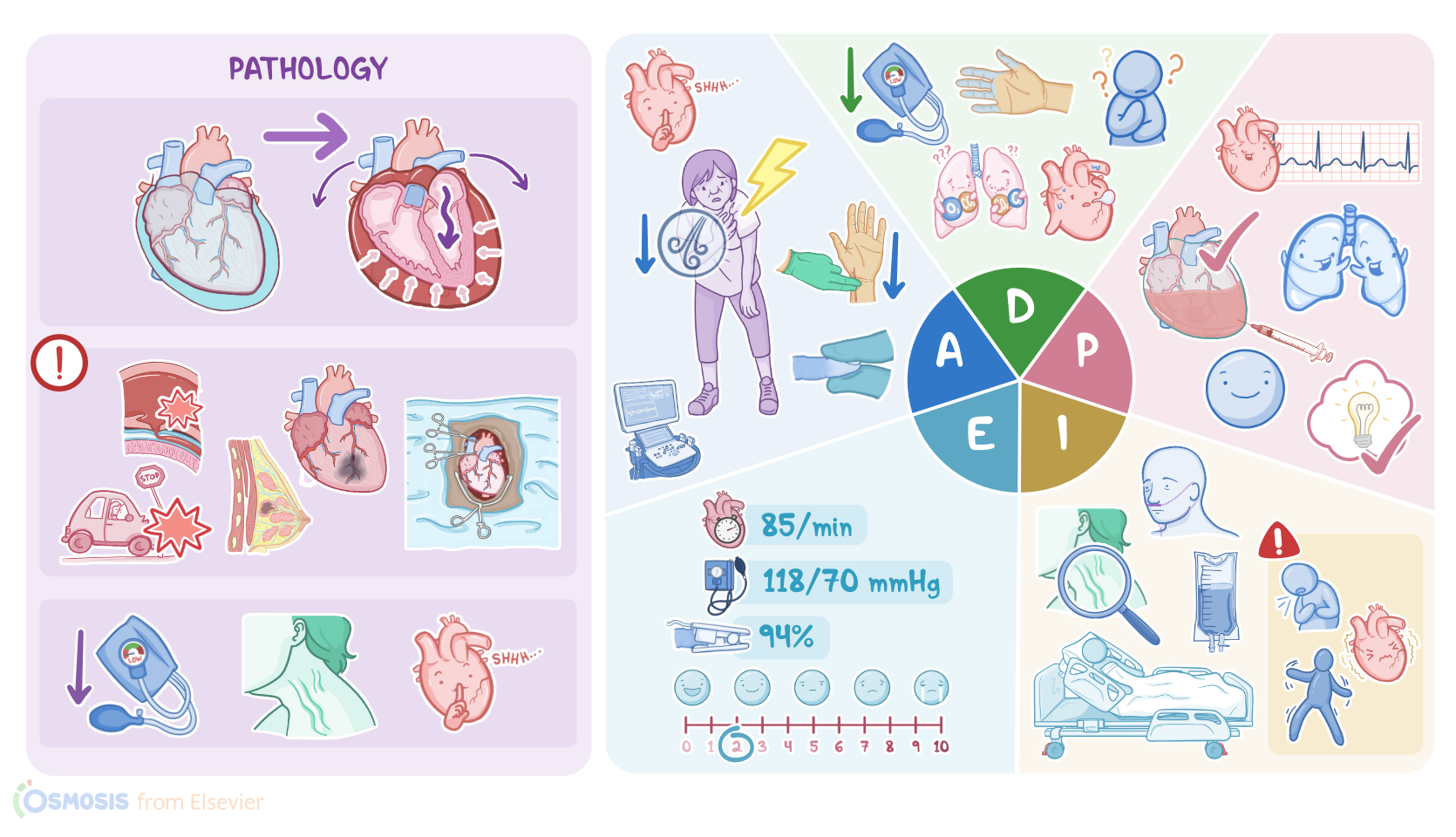
Cardiac tamponade puts pressure on the outside of the heart, restricting its movement. As a result, the heart is unable to pump normally, causing compromised cardiac functioning and resulting in decreased cardiac output.
The heart sits inside a two-layered pouch called the pericardium. Within this pouch, there's the pericardial cavity, which is filled with a small amount of fluid that lets the heart slip around as it beats.
Typically, there's approximately 15 to 50 mL of fluid in the pericardial cavity at any given time. The pericardial cavity can stretch to accommodate a gradual accumulation of fluid without immediate adverse effects.
When fluid accumulates gradually, the pericardium can hold as much as 1.5 L of fluid before tamponade sets in. In contrast, if there's a rapid buildup of fluid, even as little as 100 mL, it doesn't allow the pericardial cavity to stretch and can compress the heart, leading to cardiac tamponade.
A rapid accumulation of fluid can occur as a result of trauma to the chest. For example, a stab wound can puncture a blood vessel and fill the pericardium with blood, or blunt trauma like hitting your chest with a steering wheel during a car crash, causing the rupture of many small blood vessels.
Another cause is aortic dissection, which is when an injury to the innermost layer of the aorta allows blood to flow between the layers of the aortic wall, forcing the layers apart.
If the aortic dissection ruptures through the wall of the aorta and through the fibrous pericardium, blood can spill right into the pericardial cavity, leading to cardiac tamponade.
Cardiac tamponade can also occur a few days after a myocardial infarction or heart attack because the weak ventricular wall can rupture when exposed to the high ventricular pressure.
A rare cause is heart surgery where once again a weakened muscle can rupture and cause cardiac tamponade days after the operation.
The improper insertion of a central line or pacemaker can also increase the risk for cardiac tamponade due to the heart being perforated during the insertion or catheter migration post-insertion.
Other cases of cardiac tamponade involve long-term factors. In these cases, the fluid accumulates from a chronically inflamed pericardium that can't absorb pericardial fluid as quickly as it builds up.
Examples of this include cancers that have spread to the pericardium, such as breast or lung cancer, certain cancer treatments such as radiation therapy to the chest wall, or pericarditis, which is the inflammation of the pericardium from infection.
Cardiac tamponade has a classic clinical presentation which includes a combination of symptoms known as Beck's triad. This includes hypotension, distended neck veins, and distant or muffled heart sounds.
If cardiac tamponade develops, fluid puts pressure on the heart and prevents it from fully stretching or relaxing between contractions.
As a result, the cardiac chambers can't fill with blood properly, which causes a decrease in cardiac output leading to hypotension.
The accumulation of fluid also makes it difficult for the atria to expand enough to accommodate venous blood returning to the heart.
As a result, the blood will have nowhere to go but back into the veins. This is why you may see distended jugular veins when you look at the neck of a person with cardiac tamponade.
When listening to the chest, fluid accumulation can result in muffled or distant heart sounds. Other symptoms that can be present include pulses paradoxis, which is when the systolic BP drops more than 10 millimeters of mercury during inspiration.
Tachycardia results because less blood leaving the heart means less blood is reaching the organs and tissues, so the heart tries to compensate for the low output by beating faster.
This decreased cardiac output can also lead to weak peripheral pulses. Pressure from the fluid accumulation can also cause chest pain or discomfort.
Decreased cardiac output can cause dyspnea because there's less blood flow to the lungs. As cardiac tamponade progresses, the heart can ultimately become ischemic and stop beating altogether.
This dangerous situation requires immediate intervention. A diagnosis of cardiac tamponade often starts with an electrocardiogram or ECG.
Classic findings include sinus tachycardia, a low QRS complex voltage, and electrical alternates, which is when the QRS complexes have different heights.
However, the most precise diagnostic tool is an echocardiogram, which can show the excess pericardial fluid and the heart swinging around inside the pericardial cavity.
A chest X-ray will show an enlarged pericardial silhouette if the accumulation of fluid is more than 200 mL. Several lab tests, including complete blood count, chemistry panel, and cardiac biomarkers can be helpful in determining any contributing factors such as infection.
Cardiac tamponade is treated by inserting a needle directly into the pericardium and draining the pericardial fluid to relieve the pressure surrounding the heart, a process called pericardiocentesis.
The removal of excess fluid with a pericardiocentesis results in dramatic improvement in hemodynamic status. Prior to the procedure, the patient is provided with oxygen therapy and bed rests with the head of the bed elevated to reduce the heart's workload, IV fluids for volume expansion to increase blood flow, and medication for chest pain.
Intake and output should be monitored, in particular, urine output, since a drop in urine output may indicate decreased renal perfusion.
Following the pericardiocentesis, the underlying disease is addressed. Now that we understand pericardial effusion and cardiac tamponade, let's get back to our client, Lydia, who has now been admitted to a telemetry unit.

Assessment6:34–8:22

Her pericardiocentesis has been scheduled for the following day. After entering her room, you introduce yourself, wash your hands, and confirm her identity.
You ask her how she's doing, and she says she's anxious about her procedure tomorrow. She also states she's been having chest pain and shortness of breath the last few days.
She rates her chest pain as a 6 out of 10 on a 0 to 10 scale. You ask her if the pain radiates anywhere else, which she denies.
Upon visual inspection, you do not notice any jugular vein distention. Her lungs are clear to auscultation, but breath sounds are diminished at the bases.
Listening to her chest, her heart sounds are muffled but regular. While palpating her peripheral pulses, you make note that her pulse grade is 2+, suggesting a slightly more diminished pulse than normal.
Capillary refill is more than 2 seconds. Her skin is cool and dry.
Her vital signs are heart rate 105 BPM, BP 115/65 millimeters of mercury, respirations 24 per minute, temperature 98.6 °F or 37 °C, and oxygen saturation 90% on room air.
Her most recent diagnostic tests include an echocardiogram showing excess pericardial fluid and a chest X-ray that reveals an enlarged cardiac silhouette.
Her recent ECG shows sinus tachycardia, a low QRS complex voltage, and electrical alternatives. Her latest lab results, including a complete blood count, chemistry panel, and cardiac biomarkers, are within normal range.
Next, you document your assessment findings. Based on the assessment data you've collected, your nursing diagnoses include decreased cardiac output related to increased pericardial pressure, impaired gas exchange related to impaired ventilation and perfusion, ineffective tissue perfusion related to decreased blood flow, acute chest pain related to fluid accumulation and pericardial pressure, and deficient knowledge related to planned pericardiocentesis and post-operative care.

Diagnosis8:22–8:48

Planning8:48–9:50

After collaborating with Lydia and her healthcare team, you outlined several important goals to achieve. Lydia will maintain adequate cardiac output pre and post pericardiocentesis as evidenced by heart rate 60 to 100 BPM with regular rhythm, BP within normal range with no evidence of pulses paradoxis, and her post-procedure echocardiogram will demonstrate improved cardiac function.
Optimal gas exchange will be demonstrated by unlabored respirations at 12 to 20 per minute and oxygen saturation levels above 92%.
She'll maintain tissue perfusion as evidenced by palpation of her peripheral pulses at a grade of 3+ or a normal pulse, capillary refill under 2 seconds, and warm and dry skin.
Pain management will be demonstrated by Lydia reporting satisfactory pain control at a level less than 3 on a rating scale of 0 to 10.
Finally, she will demonstrate an understanding of the pericardiocentesis procedure and follow-up care. Now that you've established goals for Lydia, you coordinate pre-operative care with the nursing assistant and the surgical team to implement the plan of care.

Implementation9:50–11:38

Hourly vital signs are ordered, and Lydia will stay on bed rests in an upright position to relieve dyspnea and chest pain.
While completing Lydia's vital signs, you monitor for changes in cardiac rate and rhythm. You make note to look for any changes in her BP that may indicate a decline in status, including hypotension and pulses paradoxis.
Lydia's other orders include supplemental oxygen via nasal cannula to keep saturations above 92%. Next, you delegate measuring intake and output to the nursing assistant.
Lydia is ordered continuous IV fluid of 0.9% sodium chloride solution for volume resuscitation. She's also ordered IV medication, morphine, for her chest pain.
While administering her morphine, you explained the pericardiocentesis to Lydia, assuring her that she'll be monitored closely throughout the procedure and will remain in the hospital for at least 24 hours post-procedure to ensure her symptoms have resolved.
You explained that pericardial effusions can recur, and continued surveillance is important to prevent the development of cardiac tamponade.
She will need to follow up with a cardiologist once she is discharged from the hospital. You emphasize the importance of seeking medical attention for new or worsening symptoms such as pain, shortness of breath, restlessness, and palpitations.
Throughout your shift, you'll monitor closely for new or worsening symptoms that suggest cardiac tamponade. Any changes in BP or pulses paradoxis, the appearance of jugular vein distention, or changes in the ECG or echocardiogram readings will be reported to the attending physician immediately.

Evaluation11:38–13:30

A couple hours into your shift, you enter Lydia's room to evaluate how she's doing. Upon visual inspection, Lydia is anxious, short of breath, and her jugular veins appear distended.
You quickly take a set of vital signs, which are P 155 BPM, BP, 95/54 millimeters of mercury dropping to 80/54 millimeters of mercury on inspiration, respirations, 32 per minute, temperature, 98.6 °F or 37 °C, and oxygen saturation, 82% via 2 L nasal cannula.
You increase her oxygen to 4 L and notify her physician immediately. A stat, ECG and echocardiogram are completed at the bedside.
The ECG shows electrical alternans, and the echocardiogram confirms the development of cardiac tamponade. She is transferred to the cardiac catheterization lab for an emergent echocardiography guided pericardiocentesis.
The procedure is successful, and 650 mL of fluid are drained from her pericardial space. She is transferred back to the telemetry unit for at least 24 hours for continuous monitoring of her cardiac function.
Her latest vitals are P 85 BPM, BP, 118/70 millimeters of mercury, respirations, 16 per minute, temperature 98.6 °F or 37 °C, oxygen saturation, 94% on 2 L of oxygen via nasal cannula, and she's rating her pain 2 out of 10.
She will require repeat chest X-rays and echocardiograms going forward and will be followed by a cardiologist. She will follow up with her oncologist in a week to monitor her response to the procedure and make adjustments to her treatment plan as required.

Summary13:30–14:45

All right, as a quick recap, cardiac tamponade is compression on the heart that adversely affects its function caused by pericardial effusion, which is a collection of fluid inside the pericardium.
Cardiac tamponade can result from acute trauma such as a car crash or more long-term causes such as cancer. Cardiac tamponade classically presents with Beck's triad, hypotension, distended neck veins, and distant heart sounds during auscultation.
Treatment involves pericardiocentesis and addressing the underlying condition. Your assessment reveals that Lydia was experiencing shortness of breath, chest pain, muffled heart sounds, and weak peripheral pulses.
Your nursing diagnoses were decreased cardiac output, impaired gas exchange, ineffective tissue perfusion, pain, and deficient knowledge.
The goals you identified when planning care for Lydia included maintaining adequate cardiac output and tissue perfusion, improving gas exchange, controlling pain, and understanding her upcoming procedure and follow-up care.
Along with the healthcare team, you work to implement actions to achieve the goals of the plan of care for Lydia. Prior to Lydia's discharge, you and the healthcare team will continue to evaluate if these goals have been met.
Pericardial effusion and cardiac tamponade: Video | Osmosis