Placenta previa: Nursing process (ADPIE)
Client Report0:00–0:54
30-year-old Sofia Ortiz, G5 T4 P0 A0 L4, is brought to the emergency department, or ED, by her sister with painless vaginal bleeding at 34 weeks of gestation.
She says that during the last 4 weeks she has experienced intermittent spotting and that she came to the ED because the bleeding has suddenly become bright red and much heavier.
Sofia has had routine prenatal care for all of her pregnancies, and each of her babies were delivered by cesarean section.
She has no known allergies, an uncomplicated medical history, and her only medications are prenatal vitamins. A focused assessment reveals active vaginal bleeding, an absence of uterine contractions and a baseline fetal heart rate, or FHR, of 150 beats per minute.
Sofia is transferred to the labor and delivery unit for monitoring. The placenta is a temporary organ that develops in the uterus during pregnancy, and it represents a lifeline connecti on between mother and fetus.
Pathology0:54–4:32
It provides oxygen and nutrients to a growing fetus, and also removes waste products from its blood. Normally, the placenta implants in the upper uterus.
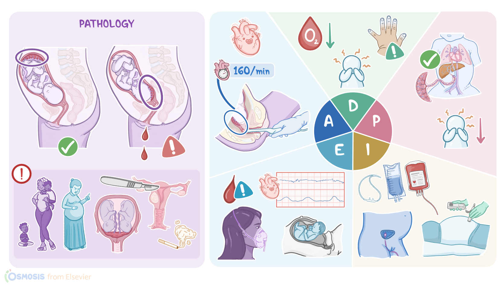
Placenta previa occurs when the placenta implants in the lower part of the uterus and partially or completely covers the opening of the cervix, referred to as the cervical os.
Now, the exact reason why placenta previa occurs is still unknown, but there are some factors that can increase the risk for it.
The first risk factor is multiple gestation, in which the uterus must accommodate either more than one fetus with a larger placenta, or more than one placenta, each containing one fetus, which increases the risk of implantation near or over the cervical os.
Additionally, abnormalities of the uterus, such as uterine fibroids, can prevent the normal implantation of an embryo. Also, previous uterine surgical interventions can cause uterine scarring and make the uterine lining less hospitable for implantation, which encourages implantation further down in the uterus.
The most important of these include previous cesarean birth and myomectomy, which is a surgical procedure used to remove uterine fibroids.
Other important procedures include multiple dilation and curettage procedures, which are performed to open the cervix and clear the uterine lining after a spontaneous or therapeutic abortion, or to remove abnormal uterine tissue.
Finally, individual risk factors include smoking, older maternal age, and multiparity, which refers to a history of two or more previous pregnancies.
Typical signs of placenta previa include sudden onset of bright red vaginal bleeding that is painless, and typically occurs after 20 weeks of gestation.
The amount of bleeding can vary, and it can increase during labor because of uterine contractions and cervical dilation.
Physical examination of clients with placenta previa reveals a soft, non-painful uterus. The fetal heart rate, or FHR, can be normal initially, but as bleeding continues over time, decreased blood flow to the placenta may result in signs of fetal hypoxia and anemia.Complications on the maternal end include hemorrhage, shock, and death.On the fetal end, placenta previa can lead to serious complications such as intrauterine growth restriction, asphyxia, preterm delivery, and death.Placenta previa is usually diagnosed with transvaginal or abdominal ultrasound, which typically reveals an abnormally positioned placenta.
A digital examination of the vagina is contraindicated if placenta previa is suspected because it can potentiate the bleeding.
Instead, examination should be performed using a speculum. In some clients, placenta previa is not recognized until cervical changes during labor cause bleeding.
For treatment, clients with placenta previa usually require hospitalization and close monitoring. The main goal is to manage bleeding, support hemodynamic stability, and to prevent preterm birth.
For minor bleeding, bed rest is usually sufficient, but for major bleeding, blood products and intravenous fluids can be provided.
In severe cases, where the mother or fetus is unstable, an immediate cesarean birth should be performed.You greet Sofia and her sister as they arrive at the Labor and Delivery unit and begin your assessment as you attach the fetal monitor, which shows an FHR of 160 beats per minute.
Assessment4:32–5:45
Variability and accelerations are present. Her uterus is soft and nontender, and active bright vaginal bleeding continues to be present on the peri-pad.
Sofia's vital signs are temporal temperature 98.6 F or 37 C, heart rate 78 beats per minute and regular, respiratory rate 16 with clear lung sounds, blood pressure 120/74 mmHg, SpO2 97% on room air, pain 0/10.
Using transvaginal ultrasonography, the midwife determines there is 3.5 cm placental tissue over the internal cervical os, consistent with a partial placenta previa.
She asks, “Will my baby be OK?” You reassure Sofia and let her know there is a team ready to help her and her baby. You document your assessment findings and develop a plan of care.
Based on your assessment findings, you determine the following priority nursing diagnoses are: risk for ineffective tissue perfusion related to blood loss; risk for ineffective fetal tissue perfusion and oxygenation related to maternal blood loss; and maternal anxiety related to threat to self and fetus.
Diagnosis5:45–6:05
After collaborating with Sofia and the midwife, you plan the following goals: Up until the time of delivery, Sofia’s hemodynamic status will be stable and she will experience decreased blood loss; there will be adequate perfusion to the fetus, and there will be normal FHR patterns; and Sofia will report that her anxiety level is manageable.
Planning6:05–6:27
Next, you implement your interventions. You review the midwife’s orders and draw blood for type and crossmatch, CBC, clotting studies, and electrolytes.
Implementation6:27–7:37
Next, you administer IV fluids, place an absorbable pad under Sofia so you can monitor blood loss, and you insert an indwelling urinary catheter.
Then, you administer oxygen at 2 L per minute via nasal cannula. You explain that her bleeding is caused by the position of the placenta, and that rest, supplemental oxygen and IV fluids will help provide the blood flow her baby needs.
As you provide care, you will closely monitor intake and output and FHR patterns, and provide Sofia with information and reassurance, while encouraging her sister’s involvement in her care as appropriate.
You will notify the midwife immediately if Sofia’s bleeding continues, if signs of hemodynamic instability are present, or if abnormal FHR patterns such as loss of variability, persistent decelerations, or sustained fetal tachycardia develop.
Let’s check back and evaluate how Sofia is doing so far. Sofia’s sister has contacted her husband, who is on his way and will arrive soon.
Evaluation7:37–8:59
Sofia is less anxious knowing her husband will arrive soon but continues to worry about her baby. Your assessment reveals continued active blood loss and these vital signs: heart rate 88 beats per minute and regular, respiratory rate 18, blood pressure 110/60 mmHg, SpO2 92% at 2 L per minute via nasal cannula, pain 0/10.
FHR is sustained at 170 bpm and a pattern of late decelerations develops. You switch Sofia’s oxygen to 8 L via mask, notify the midwife, escort Sofia to the operating suite and give report to the operative team.
Sofia’s husband arrives in time to be present at the birth. Sofia delivers a baby girl with Apgar scores of 8 and 9 and weighing 2495 g.
The neonatal team will keep a close eye on baby girl Ortiz as she transitions to extrauterine life, while the obstetric team will support Sofia until she is stable enough to be transported to the postpartum unit.
Alright, as a quick recap … Your client, Sofia Ortiz experienced painless vaginal bleeding at 34 weeks of gestation and is diagnosed with placenta previa, which is when the placenta partially or completely covers the cervix.
Summary8:59–9:44
Your nursing diagnoses included risk for ineffective tissue perfusion; risk for ineffective fetal tissue perfusion and oxygenation; and maternal anxiety.
Your planned goals and interventions focused on supporting the hemodynamic stability of Sofia and her baby and decreasing her anxiety.
While providing care you continually evaluated Sofia’s response to your interventions, and with the help of the healthcare team, you supported the safe birth of her baby girl.
you supported the safe birth of her baby girl.
| PLACENTA PREVIA | ||
| KEY POINTS | NOTES | |
| PATIENT REPORT |
| |
| PATHOPHYSIOLOGY |
| |
| DIAGNOSIS AND TREATMENT |
| |
| ASSESSMENT |
| |
| NURSING DIAGNOSES |
| |
| PLANNING |
| |
| IMPLEMENTATION |
| |
| EVALUATION |
| |
No notes for this video yet
Try adding a note below