
The 5 P's · Circulation Assessment Acronym

Published: Jan 06, 2025
Author: Lily Guo
Editor: Alyssa Haag
Editor: Józia McGowan, DO
Editor: Kelsey LaFayette, DNP, RN
Author: Jessica Reynolds, MS
Copyeditor: Stacy M Johnson, LMSW
7-day free trial

Go deeper with Osmosis

Osmosis is a learning platform with videos, questions, and AI tools to help you master topics like this.

4.8 · 12,000+ reviews
Watch quick, visual videos
Practice with Qbank-style questions
Use AI to explain, quiz, and review
Study anytime with the mobile app
Start free trial

No credit card · Cancel anytime

What is the 5 P's acronym?

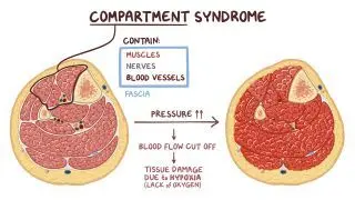
The 5 P's is an acronym used in neurovascular assessment, a routine test performed by clinicians to assess for neurovascular compromise, impaired blood flow to the extremities, and damage to the peripheral nerves. It is often used to detect compartment syndrome, a painful condition resulting from blood accumulation in an enclosed muscle space, such as in the arm or leg. Compartment syndrome warrants immediate medical treatment since it can lead to permanent muscle and nerve damage, and even death. 

Learn deeper with Osmosis

Master this topic faster with videos, questions, and AI.

Used by 8M+ healthcare learners.

Start free trial

No credit card · Cancel anytime

What are the 5 P's of circulation assessment?

The 5 P's of circulation assessment includes pain, pallor, pulse, paresthesia, and paralysis. 

Pain is assessed on a standardized scale typically from 0 to 10, with 10 being the worst pain ever experienced. Under typical conditions, pain should primarily be localized to the injury site and not be out of proportion to the severity of the injury. 

Pallor refers to the skin tone of the affected limb. Any deviation from the individual’s typical skin tone, especially blue or purple in color, can be indicative of compromised blood flow. A comparison should be made with the unaffected limb. Capillary refill time refers to the time it takes for blood to reperfuse to a body part after pressure. 

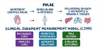
Pulse, specifically the radial, dorsalis pedis, and posterior tibial pulses, are checked to ensure proper blood flow. Depending on the source, pulses are rated on a scale of 0 to 4+, with 0 indicating no pulse and 4+ indicating a bounding pulse (i.e., strong pulsation that does not disappear with moderate pressure). A grade of 3 is considered an expected finding. 

Paresthesia refers to the sensation of numbness and tingling, which can be indicative of neurological trauma. Additionally, individuals may report diminished sensitivity to touch or feeling “pins and needles” on the affected limb. 

Lastly, paralysis refers to the inability to move the affected limb. 

How does compartment syndrome affect the 5 P's?

Compartment syndrome is a painful condition where bleeding and swelling within a limb limits blood flow and oxygen and nutrients to the muscles and nerves. This occurs due to impairment of the fascia’s ability to expand and stretch. Acute compartment syndrome is commonly due to trauma, such as a motor vehicle accident or a crush injury. Compartment syndrome of the leg is widely due to tibial shaft fracture, which requires significant force, such as from a motor vehicle accident. This is in contrast to chronic compartment syndrome, which is associated with overexertion during exercise. Severe pain is typically the first indicator of compartment syndrome. The compromised extremity may become tense and edematous due to blood accumulation, increasing the pressure on nerve fibers. This results in pain that may feel disproportionate to the visible injury. Pallor and pulselessness occur due to compromise of arterial perfusion as the limb swells, and bloody supply is occluded. Notably, it can be expected for arterial pulses and regular capillary refill to persist even in the presence of prolonged and severe compartment syndrome. 

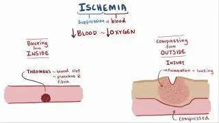
Paresthesia and complete paralysis may occur in the later stages of compartment syndrome. If the pressure in the compartment continues to increase, neuronal tissue can become ischemic, resulting in nerve dysfunction and paresthesia. In addition to feeling “pins and needles," the individual may report loss of light touch sensation, which may be assessed with a two-point discrimination test or pinprick testing. If tissue ischemia persists for more than four hours, motor function may deteriorate, resulting in paralysis of the affected limb. These functional losses may be irreversible after eight to 24 hours of tissue ischemia. In addition to feeling “pins and needles," the individual may report loss of light touch sensation, which may be assessed with a two-point discrimination test or pinprick testing. 

What are the most important facts about the 5 P's acronym?

The 5 P's acronym is used systematically in a neurovascular assessment to assess compartment syndrome's presence. The P's refer to pain, pallor, pulse, paresthesia, and paralysis. Pain is commonly rated on a 10-point scale and can be disproportionately severe in the case of compartment syndrome. Pallor refers to the appearance of the skin and whether it appears as though blood is appropriately reaching the affected body part. Signs of poor perfusion include blue or purple discoloration that only affects the compromised limb(s).

Similarly, an absent pulse indicates poor perfusion and may be experienced during the later stages of compartment pressure. Pulse is graded on a four-point scale, with three being within normal limits. Paresthesia is the presence of an abnormal sensation, such as numbness and tingling, and may occur as a symptom of nerve damage. Lastly, paralysis refers to the absence of movement and can happen if compartment syndrome has gone untreated for approximately four hours. 
Students say Osmosis is 100% worth it

Because Osmosis saves them time. Lowers stress. And actually helps them remember when it counts.

I used Osmosis to prepare for my first medical school licensing exam! Super helpful and interactive for people who may not do great with just pages of text info!

Cecilia Ruiz

Cecilia Ruiz

MD student

Sayan Misra

I have used Osmosis for about four years. Best thing I have ever used for my medical studies.

Sayan Misra

Sayan Misra

Med student

Osmosis videos are superior because they define simple concepts, tell a story with a clear progression, and provide context.

Jay Pate

Jay Pate

Dental student

References


Altizer L. Compartment syndrome. Orthopaedic Nursing. 2004;23(6):391???396. doi:https://doi.org/10.1097/00006416-200411000-00009


Andrews, LW. Neurovascular assessment. Advanced Clinical Care. 1990;5(6):5-7.


Donaldson J, Haddad B, Khan WS. The pathophysiology, diagnosis and current management of acute compartment syndrome. The Open Orthopaedics Journal. 2014;8(1):185-193. doi:https://doi.org/10.2174/1874325001408010185


Dunphy L, Morhij R, Tucker S. Rhabdomyolysis-induced compartment syndrome secondary to atorvastatin and strenuous exercise. BMJ Case Reports. Published online March 16, 2017:bcr2016218942. doi:https://doi.org/10.1136/bcr-2016-218942‌


Henry Kenneth Walker, Wilbur Dallas Hall, John Willis Hurst. Clinical Methods. Butterworth-Heinemann; 1990.


Hill RD, Smith RB. Examination of the extremities: pulses, bruits, and phlebitis. Clinics in Podiatric Medicine and Surgery. 1990;29.


Pechar J, Lyons MM. Acute compartment syndrome of the lower leg: a review. The Journal for Nurse Practitioners. 2016;12(4):265-270. doi:https://doi.org/10.1016/j.nurpra.2015.10.013


‌Rorabeck C. The treatment of compartment syndromes of the leg. The Journal of Bone and Joint Surgery British Volume. 1984;66-B(1):93-97. doi:https://doi.org/10.1302/0301-620x.66b1.6693486


Ross D. Acute compartment syndrome. Orthopaedic Nursing. 1991;10(2):33-38. doi:https://doi.org/10.1097/00006416-199103000-00007‌


Sheridan GW, Matsen FA, Krugmire RB. Further investigations on the pathophysiology of the compartmental syndrome. Clinical Orthopaedics and Related Research. 1977;&NA;(123):266???270. doi:https://doi.org/10.1097/00003086-197703000-00070


Tiwari A, Haq AI, Myint F, Hamilton G. Acute compartment syndromes. British Journal of Surgery. 2002;89(4):397-412. doi:https://doi.org/10.1046/j.0007-1323.2002.02063.x


‌Zimmerman B, Williams D. Peripheral pulse. Nih.gov. Published 2019. Accessed June 1, 2022. https://www.ncbi.nlm.nih.gov/books/NBK542175/