Physical assessment - Skin, hair, and nails: Nursing
Introduction0:00–0:22
Assessment of the skin, hair, and nails should be completed as part of a comprehensive client assessment or as a part of a focused exam when a client is experiencing issues that affect the integumentary system, such as hair loss or skin irritation.
Getting Started0:22–1:06
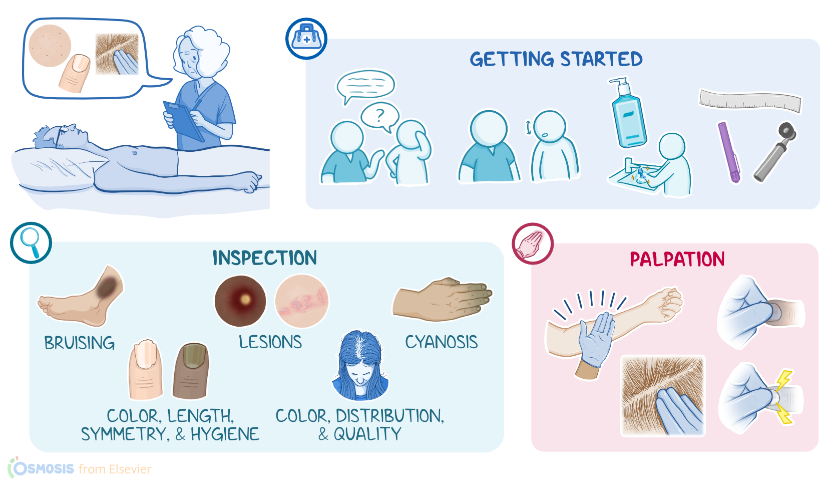
Then, prepare for the exam by ensuring your client is in a comfortable position, that your hands and stethoscope are warm, and that the temperature in the room is comfortable.
Before getting started, explain the procedure to your client and be sure to answer any questions they might have before obtaining verbal consent.
Then, perform hand hygiene and collect your supplies.There are no specific landmarks for skin and hair, but the nails do have landmarks such as the nail plate or body, lunula, cuticles, the lateral folds and the proximal nail fold.
Anatomical Landmarks1:06–1:25
Locating the anatomical landmarks of the nails will help guide your assessment. The methods of skin, hair and nails assessment include inspection and palpation.Alright, first, you should start with a general inspection of the skin, which should be intact and have a uniformity of color without areas of discoloration or the presence of lesions.
Methods of Assessment1:25–1:34
Inspection1:34–8:53
For example, calluses can appear on hands and feet due to frequent use, whereas the skin of the eyelids will be thin and delicate.
Also note your client’s skin color, which normally ranges from various shades of black, brown, and tan, to shades of white and pink.
These variations in pigmentation are due to factors like genetics and sun exposure. You should always consider these natural variations in skin color and tone when assessing your client, especially when looking for localized skin changes.
For example, let's look at rashes. In clients with light skin, they appear pinkish-red, but in clients with dark skin, look for areas of hyperpigmentation or a purplish tone.
Likewise, when assessing for bruising, in darker skin, look for deep blue or even a black tone. With lighter skin look instead for purple, blue, or even green in clients.
When assessing for a pressure injury, lighter skin will blanch, or turn white, when pressure is applied. In dark skin, however, blanching will not always occur, so you should look for areas that are shiny, indurated, or taut instead.
In clients with a more olive complexion, cyanosis can take on a gray or greenish hue. Another abnormal skin color finding is a yellow discoloration of the skin which can indicate liver disease.
This finding is often better visualized in the sclera and palms in clients with darker skin. Lastly, remember that the palms of the hands and soles of the feet are lighter in clients with darker skin, so inspection of these areas may assist in detecting widespread skin changes.
Now, some lesions you may see during inspection include macules, papules, plaques, vesicles, bullae, and pustules. First, a macule is a flat lesion, usually less than 1 centimeter wide and is a different color from the rest of the skin.
Examples include freckles and petechiae, which are tiny spots of bleeding under the skin. Next, a papule is a raised, firm demarcated lesion that’s less than 1 centimeter wide.
Examples of papules are warts and elevated nevi, or moles. Plaques are raised, firm and coarse lesions that are larger than 1 centimeter wide, as seen in clients who have psoriasis or seborrheic dermatitis.
Then there are vesicles, which are raised, superficial lesions filled with serous fluid. These are also typically less than 1 centimeter in size, and they appear in clients with chickenpox or shingles.
A bulla is a vesicle that is greater than 1 cm, and a common example is a blister. Lastly, you might visualize pustules, which are elevated lesions, similar to vesicles, but instead of serous fluid, they are filled with pus.
When direct visualization of the mole is inadequate, a penlight and magnifying glass or dermatoscope may be used.Next, inspect your client’s hair for color, distribution, and quantity which varies between each person.
Common areas for hair to exist include the scalp, neck, inside the nostrils, ears, chest, axillae, back, shoulders, upper extremities, lower extremities, pubic area, and tops of the toes.
Lack of hair on the lower extremities is an abnormal finding associated with poor perfusion. Symmetrical hair loss on the scalp commonly occurs in some clients.
However, asymmetrical hair loss is an unexpected finding, and in women, it can indicate a pathological condition such as adrenal androgenic female-pattern alopecia, which is characterized by hair thinning and loss starting at the middle of the scalp.
Now, inspection of the nails involves assessing color, length, symmetry, and hygiene. Normally, nail edges should be smooth, slightly convex, and intact; broken nails and ragged cuticles are indications of nail-biting, and peeling or nails with small pits across the nail plate are associated with psoriasis.
The nail folds should be without redness or edema. If your client reports pain, you should assess for an ingrown nail or infection.
Nail color is different for every client. The presence of streaks of color called pigment bands, is a normal finding in those with dark skin.
Yellow nails can be a sign of fungal infection or the result of nicotine staining in clients who smoke; and splinter hemorrhages may occur in clients with endocarditis or due to trauma.
The nail plate should be smooth and flat, though longitudinal ridges are an expected variation commonly associated with aging.
Transverse ridges, on the other hand, aren't expected, and can result from repeated trauma to the nail. Transverse depressions, called Beau lines, are an indication that the nail growth has been temporarily interrupted by a serious illness.Clubbing is another unexpected finding that is often related to hypoxia from chronic respiratory and cardiovascular disease.
To assess for clubbing, you can perform the Schamroth window test where your client holds two of their fingers facing opposite of each other.
Finally spooning, or a concave shape of the nails can be seen in clients with severe iron deficiency anemia.Okay, moving onto palpation.
During skin palpation, you should feel for moisture, temperature, and texture using the dorsal surface of your fingers or hand, which is more likely to detect temperature changes.
For the most part, skin should be relatively dry and contain minimal oil. Temperature of the skin varies from cool to warm, and should be symmetrical bilaterally.
Palpation8:53–10:47
If you notice unilateral temperature changes, such as one leg that’s cool and one foot that’s very warm, this can be an indication of deep vein thrombosis or cellulitis.
Overall, skin should feel smooth and even with occasional areas of dryness. A client may experience superficial hyperkeratosis, where the skin’s outer layer thickens due to overproduction of keratin, often in response to friction.
This causes areas of local scaling, roughness, calluses, or cornsTo estimate your client’s hydration status, you can assess their skin turgor.
To do this, gently pinch their skin on the forearm or under the clavicle and then release it. Normally, the skin should return to normal almost immediately.
Skin that remains tented is associated with dehydration. In older clients, skin normally remains tented due to the loss of skin elasticity.
A nail that feels too soft is associated with clubbing and a nail that’s overly thick may occur in clients with a fungal infection or shoes that are too tight.
If your assessment reveals something that’s potentially abnormal or emergent, such as a melanoma or pressure injury, you should report this immediately to the healthcare provider or dermatologist, while continuing to monitor your client’s progress or changes from baseline.Alright, as a quick recap….
Nursing Implications10:47–11:09
The methods of skin, hair and nails assessment include inspection and palpation. As the nurse, it’s your responsibility to correctly assess, interpret, report, and document your assessment findings.
Review11:09–11:42
information about the health of these parts of the body while also providing data about processes that are internal in the body The supplies you'll need for skin hair and nails assessment include a flexible ruler penlight and magnifying lens or dermato The iss of of skin hair and nails Assessment includes inspection and palpation as the nurse it's your responsibility to correctly assess interpret report and document your assessment
| PHYSICAL ASSESSMENT - SKIN, HAIR, AND NAILS | ||
| KEY POINTS | NOTES | |
| DEFINITION |
| |
| GETTING STARTED |
| |
| ANATOMICAL LANDMARKS |
| |
| METHODS OF ASSESSMENT |
| |
| INSPECTION |
| |
| PALPATION |
| |
| NURSING IMPLICATIONS |
| |
No notes for this video yet
Try adding a note below