Cataracts: Nursing
Notes
| CATARACTS | ||
| KEY POINTS | NOTES | |
| DEFINITION |
| |
| PHYSIOLOGY |
| |
| CAUSES AND RISK FACTORS |
| |
| PATHOPHYSIOLOGY |
| |
| CLINICAL MANIFESTATIONS |
| |
| DIAGNOSIS |
| |
| TREATMENT |
| |
| MANAGEMENT OF CARE |
| |
| PATIENT AND FAMILY TEACHING |
| |
Transcript
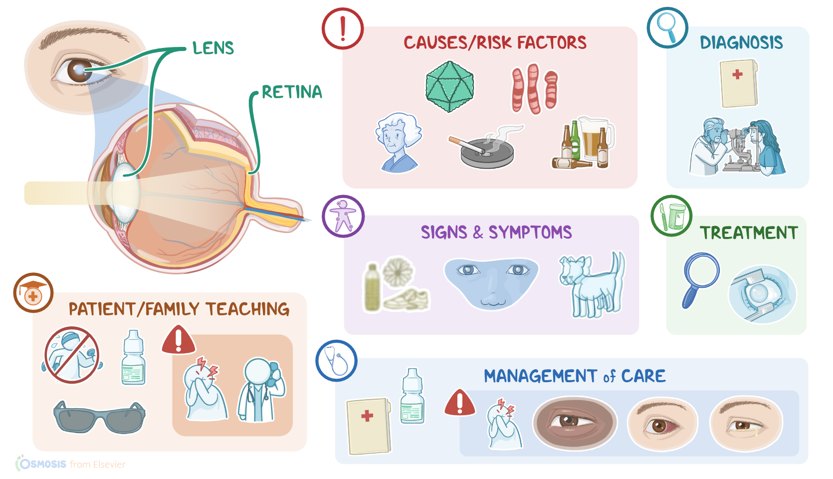
Cataracts refer to an eye condition in which the normal transparent eye lens becomes cloudy. This distorts the image projected onto the retina and causes cloudy vision.
Okay now, let’s go over some physiology. Normally, the lens is a transparent biconvex structure that lies behind the iris, dividing the anterior and posterior segments of the eye. The lens is held in place by the ciliary processes, which are tiny projections from a structure called the ciliary body. The ciliary body also controls the degree to which the lens becomes flatter or rounder. And this in turn bends the light entering the eye to focus images onto the retina. If we zoom into the lens, we’ll see that it’s composed of three layers: the capsule, cortex, and nucleus. The nucleus is made up of concentric layers of transparent proteins called crystallins.
Now, cataracts are caused by the opacification or clouding of the lens. When this occurs at birth, it's called congenital cataracts. So, risk factors for congenital cataracts include congenital infections, such as toxoplasmosis and rubella, as well as genetic conditions, such as trisomy 13, which causes Down syndrome, Wilson’s disease, in which extra copper is stored in the body tissues, and myotonic dystrophy, which is characterized by progressive muscle weakness and loss. Other risk factors for congenital cataracts include inborn errors of metabolism, like galactosemia, which is a hereditary condition that impairs the conversion of galactose to glucose in newborns.
Now, cataracts can also develop later in life, in which case it's acquired. Acquired cataracts are often associated with advanced age, usually above 60, smoking, excessive alcohol use, penetrating eye trauma, and infections. Other risk factors for acquired cataracts include exposure to UV light, prolonged use of medications like glucocorticoids, and diabetes mellitus.
Regardless of the cause, the pathological outcome is a change in the structure and composition of the lens. So, crystallins start clumping together, which causes decreased transparency of the lens, making it cloudy. At the same time, the lens becomes stiffer, which reduces its ability to refract light and focus images on the retina.
Now, the main symptom of cataracts is blurred vision that’s often bilateral, and progresses slowly over many years. This might be accompanied by decreased color perception, and diplopia or double vision. Congenital cataracts can also manifest as a gray or cloudy pupil or lack of red reflex. Diagnosis of cataracts starts with the client’s history and physical assessment, followed by slit lamp examination, which shows a loss of lens transparency.
Treatment of cataracts involves non-surgical and surgical options. Non-surgical management aims at relieving visual discomfort and might include anti-glare glasses or magnifiers, increased lighting, and lifestyle changes, such as limiting alcohol intake. Definitive treatment, though, is surgical and consists of phacoemulsification, or extracapsular extraction. In phacoemulsification, the lens is emulsified and absorbed with the help of ultrasonography, and then replaced with an artificial lens. Extracapsular extraction involves removing the lens nucleus while sparing the posterior part of the capsule to allow for the implantation of an artificial lens.
Post-operative care for clients who underwent surgical treatment of cataracts includes administering topical antibiotics and anti-inflammatory agents, in addition to analgesics and eye patches or shields.
Now, let’s look at the nursing care you would provide for a client with cataracts who has chosen to undergo a surgical revision. Your priority goals are to prepare your client for their surgery, monitor for postoperative complications, and promote safety.
Start preparing your client for surgery by reviewing their medical history and their current level of visual acuity. Take special note of the visual acuity in the unoperative eye, since they will be depending on that eye for vision while their other eye heals. Once your assessment is complete, administer the ordered preoperative medications, including nonsteroidal anti-inflammatory drops, mydriatic drops to dilate the pupil, as well as anxiolytics to reduce anxiety. Then, right before your client enters the operative suite, administer the prescribed anesthetic drops.