Anaplastic Astrocytoma · What Is It, Causes, Signs, and More
Published: Apr 07, 2026
Author: Lily Guo, MD•
Editor: Alyssa Haag, MD•
Editor: Emily Miao, MD, PharmD•
Editor: Kelsey LaFayette, DNP, ARNP, FNP-C
Illustrator: Abbey Richard, MSc
7-day free trial
Go deeper with Osmosis
Osmosis is a learning platform with videos, questions, and AI tools to help you master topics like this.
Watch quick, visual videos
Practice with Qbank-style questions
Use AI to explain, quiz, and review
Study anytime with the mobile app
Try Osmosis Free
No credit card required
What is anaplastic astrocytoma?
Anaplastic astrocytomas are rare, malignant brain tumors that arise from astrocytes, a star-shaped cell that surrounds and protects nerve cells in the central nervous system (CNS). Anaplastic refers to cancer cells that divide rapidly and do not resemble healthy cells, and astrocytes are a type of glial cell and make up the majority of cells in the human CNS. Glial cells are responsible for supporting, connecting, and protecting the neurons of the central and peripheral nervous system. Tumors arising from glial tissue, including astrocytomas, are collectively known as gliomas.
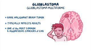
The World Health Organization (WHO) classifies astrocytomas based on how fast the cells reproduce and the likelihood of infiltration into nearby tissues. According to this classification, anaplastic astrocytomas are grade III and are considered high-grade (i.e., degree of abnormality among cells when viewed microscopically). This is in contrast with grade I or II astrocytomas which are circumscribed and low-grade, or grade IV astrocytomas which are also known as glioblastoma multiforme (GBM), which are the most aggressive form of astrocytomas. Low-grade astrocytomas can transform into high-grade astrocytomas over time.
Learn deeper with Osmosis
Master this topic faster with videos, questions, and AI.
Used by 8M+ healthcare learners.
Try Osmosis Free
No credit card required
What causes anaplastic astrocytoma?
The exact cause of anaplastic astrocytoma is unknown, however, it is thought to arise from a combination of genetic and environmental factors. Genetic mutations in astrocytes can lead to uncontrolled cell growth which underlies the pathophysiology of anaplastic astrocytomas. Additionally, mutations in tumor suppressor genes (e.g., TP53) can increase genetic susceptibility to astrocytomas. Environmental factors may also trigger these genetic changes, including exposure to ultraviolet rays, chemicals, and ionizing radiation, as well as diet (e.g., cured meats that contain high levels of N-nitroso compounds [NOCs]) and stress. A general risk factor for developing brain tumors includes a family history of brain tumors. Additionally, astrocytomas occur at a greater frequency in those with certain genetic disorders (e.g., neurofibromatosis type I, Li-Fraumeni syndrome, tuberous sclerosis).
Anaplastic astrocytomas affect genetic males more than genetic females and are more common in adults compared to children. The average age is typically between 30 and 50 years, whereas in children, they develop between 5 and 9 years of age.
What are the signs and symptoms of anaplastic astrocytoma?
The signs and symptoms of anaplastic astrocytoma occur due to the effects of the tumor or mass on the surrounding areas of the brain, and the increased intracranial pressure caused by the tumor. Typically, anaplastic astrocytomas grow slowly over time, but they may develop rapidly resulting in a sudden onset of symptoms. Generally, symptoms may include headaches that are often worse in the morning, altered mental status, lethargy or drowsiness, nausea, vomiting, and seizures. Anaplastic astrocytomas can develop anywhere in the central nervous system, however, most are found in the cerebrum (i.e., the largest part of the brain) which is divided into two hemispheres and four lobes (i.e., frontal, temporal, parietal, and occipital). If the frontal lobe is affected, the individual may experience changes in mood and personality, difficulties with memory, and paralysis of the opposite side of the body from where the tumor is located. If the temporal lobe is affected, the individual may experience problems with coordination and speech, as well as seizures. Parietal lobe tumors can lead to sensory abnormalities including tingling or burning sensations, trouble with fine motor coordination, and difficulty reading and writing. Lastly, tumors in the occipital lobe can result in vision changes or vision loss.
How is anaplastic astrocytoma diagnosed?
Anaplastic astrocytomas can be diagnosed via a thorough clinical evaluation, including a detailed patient history and physical examination. If a brain tumor is suspected after clinical examination, imaging is oftentimes ordered to evaluate the size, location, and spread of the tumor, which can also aid with surgical planning. Magnetic resonance imaging (MRI) is the most useful for visualizing soft tissue masses such as brain tumors; however, computed tomography (CT) scans may also be used. On MRI, an astrocytoma may appear as an inhomogeneous lesion with perifocal edema, without necrosis. A biopsy of the tumor, whereby a sample of tissue is taken and examined under the microscope, can provide a definitive diagnosis and determine the type of malignancy as well as tumor grade.
How is anaplastic astrocytoma treated?
Treatment of anaplastic astrocytomas frequently involves a team of specialists, which may consist of an oncologist, radiation oncologist, neurosurgeon, neurologist, nurses, and other healthcare professionals. The specific therapies utilized can vary depending on the size, location, grade, and stage (i.e., the spread or infiltration) of the tumor.
Initial treatment options for anaplastic astrocytoma typically consist of surgical excision and resection, or removal of the bulk of the tumor to preserve and improve quality of life. If the tumor cells have spread into surrounding brain tissue, radiation can be used to treat possible residual cancer. If surgery is not an option due to factors such as the location of the tumor and the individual’s age, radiation therapy alone may be used. Chemotherapy with anticancer drugs such as temozolomide can be used in conjunction with radiation to treat anaplastic astrocytoma. Temozolomide is the only drug approved by the Food and Drug Administration (FDA) for treatment in adults with refractory anaplastic astrocytoma, which has not responded to other forms of therapy. At this time, there are no chemotherapeutic agents approved for the treatment of children with anaplastic astrocytoma, however, clinical trials and experimental therapies are currently underway.
What are the most important facts to know about anaplastic astrocytoma?
Anaplastic astrocytomas are malignant, high-grade brain tumors that arise from astrocytes, a type of cell that supports and protects the brain's neurons. The exact cause of anaplastic astrocytoma is unknown, however, it is thought to arise from a combination of genetic mutations and environmental factors. Symptoms can vary depending on the tumor's location and generally include headaches, lethargy, vomiting, seizures, and changes in mood, personality, coordination, sensation, and vision. Diagnosis involves a thorough clinical evaluation, imaging tests like MRI or CT scans, and a biopsy to determine the tumor's malignancy and grade. Treatment typically requires a multidisciplinary approach, including surgery to remove as much of the tumor as possible, followed by radiation and possibly chemotherapy, especially if the tumor is inoperable or has spread.
Key Takeaways
Definition | Anaplastic astrocytomas are rare, malignant brain tumors that arise from astrocytes, a star-shaped glial cell that surrounds and protects nerve cells in the central nervous system (CNS). |
- Support, connect, and protect neurons of the central and peripheral nervous system - Gliomas = tumors arising from glial tissue | |
WHO Astrocytoma Classification | - Grade I and II → circumscribed and low-grade - Grade III → anaplastic astrocytoma (high-grade) - Grade IV → glioblastoma multiforme (GBM) (high-grade, most aggressive) *Possible transformation low-grade into high-grade |
Causes | - Genetic mutations → uncontrolled growth - Environmental risk factors triggering genetic changes - Exposure to UV rays, chemicals, ionizing radiation - Diet - Stress - Other risk factors: - Family history of brain tumors - Predisposing genetic disorders: neurofibromatosis type I, Li-Fraumeni syndrome, tuberous sclerosis - Population: - Genetic males > genetic females - Adults > children - Average age: 30-50 years in adults, 5-9 years in children |
Signs and Symptoms | Mass effect on surrounding brain areas + increased intracranial pressure | can develop rapidly - Headaches (worse in the morning) - Altered mental status - Lethargy - Nausea and vomiting - Seizures - Depending on location: - Frontal lobe → mood/personality changes, memory problems, contralateral hemiparalysis - Temporal lobe → coordination and speech impairment, seizures - Parietal lobe → sensory abnormalities, fine motor coordination impairment, reading and writing impairment - Occipital lobe → vision changes or vision loss |
Diagnosis | - Medical history - Physical examination - MRI or CT scan - Tumor biopsy |
Treatment | - Multidisciplinary team - Treatment options: - Surgical excision - Radiation therapy (after surgery or alone, if surgery not feasible) +/– chemotherapy (adults) |
Related topics

Students say Osmosis is 100% worth it
Because Osmosis saves them time. Lowers stress. And actually helps them remember when it counts.
I used Osmosis to prepare for my first medical school licensing exam! Super helpful and interactive for people who may not do great with just pages of text info!"

Cecilia Ruiz
MD student

I have used Osmosis for about four years. Best thing I have ever used for my medical studies."

Sayan Misra
Med student
Osmosis videos are superior because they define simple concepts, tell a story with a clear progression, and provide context."

Jay Pate
Dental student
References
Grimm SA, Chamberlain MC. Anaplastic astrocytoma. CNS Oncol. 2016;5(3):145-157. doi:10.2217/cns-2016-0002
Louis DN, Ohgaki H, Wiestler OD, et al. The 2007 WHO classification of tumours of the central nervous system. Acta Neuropathol. 2007;114(2):97–109.
National Organization for Rare Disorders. Anaplastic Astrocytoma - Symptoms, Causes, Treatment. rarediseases.org. Accessed September 2, 2025. https://rarediseases.org/rare-diseases/anaplastic-astrocytoma/#symptoms
UCSF Brain Tumor Center. Anaplastic astrocytoma (grade III). braintumorcenter.ucsf.edu. Accessed September 2, 2025. https://braintumorcenter.ucsf.edu/condition/anaplastic-astrocytoma-grade-iii