Eschar · What It Is, Causes, Treatment, and More

Published: Sep 29, 2025
Author: Katie Arps, BSN, RN
Editor: Alyssa Haag, MD
Editor: Emily Miao, PharmD, MD
Editor: Kelsey LaFayette, DNP, ARNP, FNP-C
Editor: Lily Guo, MD
Illustrator: Jessica Reynolds, MS
Copyeditor: Sadia Zaman, MBBS, BSc
7-day free trial

Go deeper with Osmosis

Osmosis is a learning platform with videos, questions, and AI tools to help you master topics like this.

4.8 · 12,000+ reviews
Watch quick, visual videos
Practice with Qbank-style questions
Use AI to explain, quiz, and review
Study anytime with the mobile app
Start free trial

No credit card · Cancel anytime

What is eschar?

Eschar refers to necrotic, or dead, tissue that can develop on severe wounds. It’s typically dry, black, firm, and adhered to the wound bed and edges. Eschar can occur on full-thickness injuries, which are wounds that extend below the epidermis and dermis. Examples include third degree burns or stage three and four pressure injuries. Additionally, eschar can be present on some skin rashes associated with infections, such as ecthyma gangrenosum, scrub typhus, rickettsialpox, and anthrax 

Eschar differs from a scab, which forms when platelets and fibrinogen form a fibrin mesh, trap red blood cells on surface wounds, and form a clot that dries into a scab. In contrast, eschar forms when dead tissue debris from a full-thickness wound dries out and hardens.  

Learn deeper with Osmosis

Master this topic faster with videos, questions, and AI.

Used by 8M+ healthcare learners.

Start free trial

No credit card · Cancel anytime

Should eschar be removed?

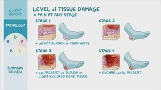
Eschar is generally removed since the necrotic tissue often impedes wound healing.   Tissue death inactivates growth factors which normally help in tissue regeneration and slows the formation of granulation tissue in the wound bed. Additionally, when dead tissue dries out and becomes hardened eschar, it physically obstructs wound contraction and stunts epithelialization, a stage in wound healing.  

Debriding eschar early in the healing process can shorten healing time. A notable exception to early debridement could be a stable eschar (i.e., an eschar that is dry, adherent, and intact) that is present on a heel or ischemic limb. Due to inherent decreased blood flow to these areas, some experts argue that the benefit of leaving the eschar in place to reduce the risk of infection outweighs the benefits of debriding. 

In certain types of wounds, such as circumferential burns, constriction caused by the presence of eschar can  cause compartment syndrome, which is an increase in pressure within a muscle compartment potentially leading to muscle and nerve damage. Sharp debridement with a scalpel may be difficult due to the firm, dry nature of eschar. Other techniques include surgical, enzymatic/chemical, mechanical, and biological debridement. During surgical debridement, the individual is usually placed under anesthesia to remove dead tissue. Enzymatic, or chemical debridement involves use of a topical enzyme to break down the necrotic tissue . Mechanical debridement can be performed with wet-to-dry dressing designed to soften and remove the eschar.  Other mechanical options include whirlpool baths or pulse lavage. Lastly, biological debridement involves the use of sterile maggots to eat away at the dead tissue, while preserving live tissue.  

Key Takeaways

Definition

Eschar refers to necrotic tissue that can develop on severe wounds, appearing as dry, black, firm, and adherent to the wound bed and edges. It occurs on full-thickness injuries, such as third-degree burns, or in association with some skin rashes, such as ecthyma gangrenosum, scrub typhus, rickettsialpox, and anthrax 

Removal 

- Removal is generally recommended  

- Necrotic tissue impairs wound healing by: 

     - Inactivating growth factors needed for tissue regeneration 

     - Obstructing wound contraction and epithelialization 

     - Early debridement can shorten healing time 

     - Exception: stable eschar on heel or ischemic limb 

- In certain wounds (e.g., circumferential burns), eschar can cause constriction → risk of compartment syndrome  

- Debridement techniques:  

     - Sharp debridement (scalpel) 

     - Surgical debridement  

     - Enzymatic/chemical debridement   

     - Mechanical debridement 

     - Wet-to-dry dressings  

     - Whirlpool baths 

     - Pulse lavage 

     - Biological debridement (sterile maggots) 

Students say Osmosis is 100% worth it

Because Osmosis saves them time. Lowers stress. And actually helps them remember when it counts.

I used Osmosis to prepare for my first medical school licensing exam! Super helpful and interactive for people who may not do great with just pages of text info!

Cecilia Ruiz

Cecilia Ruiz

MD student

Sayan Misra

I have used Osmosis for about four years. Best thing I have ever used for my medical studies.

Sayan Misra

Sayan Misra

Med student

Osmosis videos are superior because they define simple concepts, tell a story with a clear progression, and provide context.

Jay Pate

Jay Pate

Dental student

References


DeMarco S. Wound and pressure ulcer management. Johns Hopkins Medicine. https://www.hopkinsmedicine.org/gec/series/wound_care.html. Accessed August 10, 2022.


Ennis WJ, Hill D. Wound healing: A comprehensive wound assessment and treatment approach. In: Albanna MZ, Holmes JH, eds. Skin Tissue Engineering and Regenerative Medicine. Elsevier; 2016:239-263.


Franklin Institute. All about scabs. The Franklin Institute. https://www.fi.edu/heart/all-about-scabs. Accessed August 5, 2022.


NHS. Compartment syndrome. NHS. https://www.nhs.uk/conditions/compartment-syndrome/. Accessed August 15, 2022.


Silvestri LA. Integumentary system. In: Silvestri AE, ed. Saunders Comprehensive Review for the NCLEX-RN Examination. 7th ed. St Louis: Elsevier; 2017:544-568.


Walesko MB, Javier NM. Pressure injury. In: Ferri FF, ed. Ferri's Clinical Advisor 2022. Elsevier; 2022:1249-1253.