Jaw claudication can be difficult to diagnose as its symptoms may resemble those of many other conditions. Diagnosis typically begins by taking a thorough medical history and conducting a thorough physical exam. The physical exam should focus on the temporal arteries, which may feel tender, have a reduced pulse, or have a hard, cordlike feel and appearance. Additionally, fundoscopic examination might show optic disc pallor and edema early in the disease, and in more advanced cases, there may even be optic disc atrophy.
Certain blood tests may also aid in the diagnosis of jaw claudication and its underlying cause. For example, erythrocyte sedimentation rate (ESR) and C-reactive protein (CRP) are two blood markers that are elevated when there is inflammation in the body, such as in temporal arteritis. A Doppler ultrasound may be performed to identify signs of inflammation and estimate blood flow through the temporal arteries, while other imaging tests, like magnetic resonance angiography or positron emission tomography (PET) may be useful to obtain detailed images of the affected blood vessels and highlight areas of inflammation, respectively.
Lastly, the gold standard method of diagnosing temporal arteritis is by taking a temporal artery biopsy. The biopsy may show granulomatous inflammation with abnormally large cells, called giant cells. However, many individuals with temporal arteritis have a negative or unconclusive temporal artery biopsy. That is usually because temporal arteritis affects the artery segmentally, so a small tissue sample may lack pathologic findings, leading to false-negative biopsy results. Literature reviews propose that a negative temporal artery biopsy should not change the management of temporal arteritis if the clinical suspicion is high, and that non-invasive imaging modalities may be sufficient to confirm the diagnosis in most cases.
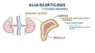
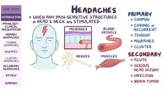
Treatment of jaw claudication depends on the underlying cause. In the case of temporal arteritis, treatment primarily consists of high dose corticosteroids, such as prednisone. Once acute symptoms resolve, the dose of corticosteroids is gradually reduced to the lowest effective dose over the course of a few months. During this period, some symptoms such as headaches may return, which can be treated with slight increases in the corticosteroid dose.
Since high dose corticosteroids can lead to significant side effects (e.g., weakened bones, high blood pressure and blood sugar, mood swings, etc.), it is important to monitor the individual's bone density and to initiate prompt calcium and vitamin D supplementation to prevent bone loss. Alternative therapy options for temporal arteritis include immunosuppressant drugs, such as methotrexate or tocilizumab. These medications are used as steroid-sparing agents, as they can help lower the individual’s steroid dosage or taper the steroids more rapidly, which is helpful for people with certain conditions like diabetes and osteoporosis.