Pyoderma Gangrenosum · What It Is, Causes, Signs, Symptoms, and More

Published: Jun 01, 2026
Author: Georgina Tiarks
Editor: Alyssa Haag, MD
Editor: Stefan Stoisavljevic, MD
Editor: Kelsey LaFayette, DNP, ARNP, FNP-C
Illustrator: Jessica Reynolds, MS
7-day free trial

Go deeper with Osmosis

Osmosis is a learning platform with videos, questions, and AI tools to help you master topics like this.

4.8 · 12,000+ reviews
Watch quick, visual videos
Practice with Qbank-style questions
Use AI to explain, quiz, and review
Study anytime with the mobile app
Start free trial

No credit card · Cancel anytime

What is pyoderma gangrenosum?

Pyoderma gangrenosum (PG) is a painful, rapidly progressive ulcerative skin condition characterized by dysfunctional neutrophils. It is a rare disorder that can appear anytime; however, it is most commonly seen in individuals between their 40s and 50s. Due to the severe pain caused by PG, it can be a distressing disease that affects an individual's activities of daily living. 

Learn deeper with Osmosis

Master this topic faster with videos, questions, and AI.

Used by 8M+ healthcare learners.

Start free trial

No credit card · Cancel anytime

What causes pyoderma gangrenosum?

While the exact cause is not known, pyoderma gangrenosum is thought to be associated with an impaired inflammatory response. Researchers have found several important factors involved in this condition that support this association, including neutrophil dysfunction, increased cytokine response, and a possible genetic component. Neutrophil dysfunction includes abnormal neutrophil chemotaxis (i.e., neutrophil recruitment), migration, phagocytosis (i.e., engulfment), and bactericidal properties. The increased cytokine response may involve the overexpression of inflammatory mediators such as TNF-alpha, chemokines, metalloproteinases, and IL-1. Lastly, deficiencies of the Janus kinase (JAK) pathway, which is involved in the immune system, have also been proposed. 

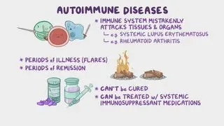
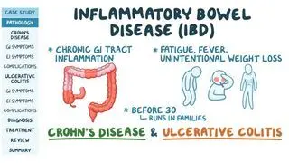
The onset of pyoderma gangrenosum has been linked to various inflammatory conditions and malignancies. Most commonly, PG is associated with inflammatory bowel disease (i.e., ulcerative colitis and Crohn disease). There are arthritic diseases related to PG, such as psoriatic arthritis (i.e., joint and skin inflammation); polyarthritis (i.e., joint disease of more than five different joints); spondyloarthritis (i.e., arthritis in spine); and rheumatoid arthritis (i.e., an autoimmune disease affecting joints). Additionally, hepatitis (i.e., inflammation of the liver) and primary biliary cirrhosis (i.e., autoimmune disease of bile ducts) are diseases affecting the liver and biliary system that can be associated with PG. Finally, in rare occasions, autoimmune conditions such as systemic lupus erythematosus (i.e., widespread inflammation impacting tissue all around the body) and Sjogren syndrome (i.e., dry eyes and dry mouth) may also co-occur with PG.  
What are the signs and symptoms of pyoderma gangrenosum?

The signs and symptoms of pyoderma gangrenosum usually include the development of painful, erythematous (i.e., red) ulcerations with a violaceous (i.e., violet) border. Initially, mild trauma to the skin may develop into a pustule or papule that looks like an insect bite. Over time, the inner wound may become exudative, while the outer edges may become erythematous and violaceous. Over time, the inner bed may also appear necrotic with a black eschar. A few weeks after the initial development of the wound, granulation tissue may appear, and a cribriform scar (i.e., uneven scarring) can grow. These ulcerations appear most often on the lower extremities; however, they may occur anywhere on the body, including the genitals and eyes. In addition to the ulcerative wound, an individual might experience arthralgias (i.e., joint pain) and fatigue. Pathergy may also occur when mild trauma results in excessive epidermal destruction, thereby causing an enlarging wound that is out of proportion to the initial incident. 

How is pyoderma gangrenosum diagnosed?

Pyoderma gangrenosum may be diagnosed after a thorough medical history and physical examination. A detailed review of systems is crucial to uncover any other systemic conditions accompanying the PG. PG is a diagnosis of exclusion, meaning that all other possible causes must be ruled out before a conclusion may be reached. 

A provider may want to order various tests to rule out other causes or look for a coinciding systemic illness. A complete blood count (CBC) assesses for anemia and elevated white blood cell count indicating inflammation. A comprehensive metabolic panel (CMP) can determine electrolyte levels, kidney function, and liver function. To look further into blood disorders, serum electrophoresis, blood smear, and bone marrow biopsy may be performed to look for hematologic causes. Antibodies such as an antineutrophil cytoplasmic antibody (ANCA), antinuclear antibody (ANA), and antiphospholipid antibody may be used to rule out other autoimmune conditions.  Tissue cultures and biopsies may be performed to rule out infections (e.g., fungal, bacterial, viral) or malignancy. An ulcer biopsy can show neutrophilic infiltration and necrosis of the epidermis. If an individual is experiencing any gastrointestinal symptoms, a colonoscopy may be considered to assess for inflammatory bowel disease. 

Researchers have recently proposed guidelines to aid healthcare providers in diagnosing PG. Major criteria that must be present include progressive ulcer and excluding all other conditions.  Additionally, four minor criteria might be found, including pathergy or cribriform scarring;  having other coinciding systemic diseases; typical biopsy findings;  and a response to treatment with steroids. 

How is pyoderma gangrenosum treated?

Pyoderma gangrenosum may be treated through various mechanisms based on the severity and response to therapy. Dermatologists may be employed to manage and oversee the treatment plan. Initially, wound care includes cleaning the site and using bandages to secure the area. Some health care providers may consider debridement; however, recent discussions have suggested debridement may increase the risk of pathergy. Topical treatment includes corticosteroids and immunosuppressants like tacrolimus, which may be used to reduce inflammation at the site. However, if the condition is progressing quickly or not responding to local treatment, systemic treatment, such as prednisone, may be used. Additionally, systemic immunosuppressants (i.e., cyclosporine, azathioprine, cyclophosphamide) and TNF-alpha inhibitors (i.e., etanercept, infliximab) may also be used independently or added to the treatment. If a quick response is needed, intravenous methylprednisolone and an immunosuppressant might be used. Hyperbaric oxygen can also be used as an adjuvant therapy to accelerate wound healing. 

What are the most important facts to know about pyoderma gangrenosum?

Pyoderma gangrenosum is neutrophilic dermatitis characterized by painful, erythematous ulcerative wounds. While the exact cause of PG is unclear, the immune system plays a significant role, specifically dysfunctional neutrophil chemotaxis and migration. PG has also been associated with multiple systemic conditions, including autoimmune disorders, arthritic disease, inflammatory bowel disease, and hematologic involvement. Individuals with pyoderma gangrenosum usually develop multiple rapidly progressive ulcerative wounds. They may also experience pathergy due to tissue destruction. Diagnosis is based on the exclusion of other possible underlying causes. Treatment for pyoderma gangrenosum is complex. Initially, topical immunosuppressants may be used, but systemic corticosteroids and immunosuppressants can be employed if necessary. 
Students say Osmosis is 100% worth it

Because Osmosis saves them time. Lowers stress. And actually helps them remember when it counts.

I used Osmosis to prepare for my first medical school licensing exam! Super helpful and interactive for people who may not do great with just pages of text info!

Cecilia Ruiz

Cecilia Ruiz

MD student

Sayan Misra

I have used Osmosis for about four years. Best thing I have ever used for my medical studies.

Sayan Misra

Sayan Misra

Med student

Osmosis videos are superior because they define simple concepts, tell a story with a clear progression, and provide context.

Jay Pate

Jay Pate

Dental student

References


Barbe, M., Batra, A., Golding, S., et al. Pyoderma gangrenosum: A literature review. Clin Podiatr Med Surg. 2021;38(4):577-588. doi:10.1016/j.cpm.2021.06.002 


George, Christina, et al. “Pyoderma gangrenosum – A guide to diagnosis and management.” Clinical Medicine, vol. 19, no. 3, May 2019, pp. 224–28. PubMed Central, https://doi.org/10.7861/clinmedicine.19-3-224. 


Maverakis, E., Marzano AV, Le ST, et al. Pyoderma gangrenosum. Nat Rev Dis Primers. 2020;6(1):81. doi:10.1038/s41572-020-0213-x