Skin Lesions · What Are They, Types, Causes, Diagnosis, Treatment, and More
Published: Mar 04, 2025
Author: Anna Hernández Castillo, MD•
Editor: Antonella Melani, MD•
Editor: Lisa Miklush, PhD, RN, CNS
Illustrator: Abbey Richard
7-day free trial
Go deeper with Osmosis
Osmosis is a learning platform with videos, questions, and AI tools to help you master topics like this.
Watch quick, visual videos
Practice with Qbank-style questions
Use AI to explain, quiz, and review
Study anytime with the mobile app
Try Osmosis Free
No credit card required
What is a skin lesion?
A skin lesion refers to any skin area that has different characteristics from the surrounding skin, including color, shape, size, and texture. Skin lesions are very common and often appear as a result of a localized damage to the skin, like sunburns or contact dermatitis. Others, however, can be manifestations of underlying disorders, such as infections, diabetes, and autoimmune or genetic disorders. Although most skin lesions are benign and harmless, some of them can be malignant or premalignant, meaning they have the potential to evolve into skin cancer.
Learn deeper with Osmosis
Master this topic faster with videos, questions, and AI.
Used by 8M+ healthcare learners.
Try Osmosis Free
No credit card required
What is a primary skin lesion?
Skin lesions can be divided into two main types: primary and secondary. Primary skin lesions originate on previously healthy skin and are directly associated with a specific cause. Common examples of primary skin lesions include freckles, moles, and blisters, among others. On the other hand, secondary skin lesions develop from the evolution of a primary skin lesion, either due to traumatic manipulation, such as scratching or rubbing, or due to its treatment or progression. Examples of secondary skin lesions include crusts, sores, ulcers, and scars.
What do skin lesions look like?
Skin lesions can present in a variety of different sizes, shapes, and forms. Moreover, skin lesions can appear isolated or in groups, and either localized in a single area or widespread throughout the body.
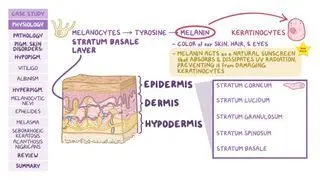
Macules are flat, well-circumcised lesions up to 1 cm (0.39 inches) in diameter, while patches are similar but are larger than 1 cm. Papules are raised bumps that are up to 1 cm in diameter; plaques are similar, though larger than 1 cm. A smooth papule or plaque that is transient (meaning that it occasionally appears and disappears) is called a wheal. Vesicles (such as with herpes simplex infections) are up to 1 cm in diameter and look like clear, fluid-filled blisters, while bullae are larger than 1 cm. Pustules (such as pimples or acne) are pus-filled, elevated lesions up to 1 cm in diameter. Finally, scales are accumulations of thickened stratum corneum (the outermost layer of the skin, consisting of dead skin cells) that become dry and flaky and sometimes peel off; while crusts are dry exudates like sebum, pus, or blood.What is a malignant skin lesion?
A malignant skin lesion is, by definition, skin cancer. The two main types of skin cancer are keratinocyte carcinoma and melanoma. Each type of skin cancer has unique characteristics, but general signs of skin cancer can include rapidly growing skin lesions, changes in the color or size of a preexisting lesion, or a scabbing sore that doesn’t heal with time.
What do malignant skin lesions look like?
Keratinocyte carcinoma arises from skin cells called keratinocytes, and includes basal cell carcinoma and squamous cell carcinoma. Basal cell carcinoma can appear as a pearly, flesh-colored skin lesion, with superficial blood vessels called telangiectasias on top. Basal cell carcinoma may present as a superficial scaling plaque, or a non-healing sore, which may bleed or form a crust. Conversely, squamous cell carcinoma commonly appears as a thick, crusty sore, with a reddish, inflamed base that can ulcer (appear as an open sore) and bleed.
Melanoma arises from skin cells called melanocytes. Melanoma typically looks like an abnormal or irregular mole. The main warning signs of melanoma can be assessed using the ABCDE rule. ABCDE stands for Asymmetry, Border irregularities, Color heterogeneity, Diameter over 6 mm, and Evolution, which refers to changes in size, color, or shape over time. The presence of one or more of these features indicates a higher chance of malignancy.
What is a benign skin lesion?
A benign skin lesion is a non-cancerous skin abnormality, growth, or tumor that can occur anywhere on the body. Benign lesions can manifest in a number of different ways, depending on their cause and tissue of origin. Common benign skin lesions include most melanocytic nevi, better known as moles, seborrheic keratoses, skin tags, cherry angiomas, and lipomas, among others. Most of the time, these lesions are harmless and don’t require treatment, unless they cause symptoms such as discomfort or itching.
What do benign skin lesions look like?
Unlike malignant lesions, benign skin lesions are generally symmetrical, well-circumscribed, have a uniform appearance, and are stable or grow slowly over time. However, in certain cases, it can be difficult to distinguish between benign and malignant lesions; in those cases, a biopsy or surgical removal of the affected area can be performed to rule out malignancy.
Benign lesions should also be distinguished from premalignant lesions such actinic keratosis or lentigo maligna, which present an increased risk of developing into different types of skin cancer. Both actinic keratosis and lentigo maligna occur as a result of long term unprotected sun exposure. Actinic keratosis appears as dry, scaly patches of skin over sun-exposed areas, like the nose and forehead, whereas lentigo maligna takes the appearance of localized dark-brown or black lesions, predominantly on the face and trunk.What causes skin lesions?
Skin lesions can be hereditary (for example, moles or birthmarks), or acquired as a result of a variety of conditions. One of the most common causes of skin lesions are infections; these include viruses, like herpes simplex, human immunodeficiency virus (HIV) or human papillomavirus (HPV); bacteria, such as Staphylococcus or Streptococcus species; and fungi like Candida albicans. Other causes of skin lesions include allergic reactions; side effects of certain medications, like corticosteroids or chemotherapy; contact with irritant substances; unprotected sun exposure; severe burns; insect bites; poor circulation; vitamin deficiencies; systemic diseases like autoimmune diseases; some infectious diseases; liver and kidney disease; and cancer.
Can cancer cause skin lesions?
Cancer can cause skin lesions through the spreading of malignant cells to the skin or, more commonly, as a result of paraneoplastic syndromes, which are distant clinical manifestations triggered by an internal malignancy. Examples of cutaneous paraneoplastic syndromes include dermatomyositis (which causes skin rashes and muscle weakness) and pyoderma gangrenosum (which is a rapidly enlarging, painful ulcer). Other paraneoplastic conditions that involve the skin include Sweet’s syndrome, which causes skin lesions along with sudden onset fever, and the Leser–Trélat sign, which includes the presence of multiple seborrheic keratoses.
Does AIDS cause skin lesions?
Individuals with AIDS can develop skin lesions as a result of the progressive immunosuppression caused by HIV infection. AIDS is associated with Kaposi sarcoma, an aggressive type of cancer that presents as red or purplish nodules and plaques on the skin and mucous membranes, such as the gums. Besides Kaposi sarcoma, individuals with HIV infection can develop severe opportunistic skin infections, as well as skin rashes (such as Stevens–Johnson syndrome) as a side effect of antiretroviral treatment.
Stevens–Johnson syndrome is a severe and potentially fatal skin condition that affects the skin and mucous membranes, such as the eyes, mouth, and genitals, causing extensive rashes and painful blisters that rapidly spread throughout the body. Most cases of Stevens–Johnson syndrome are preceded by flu-like symptoms, such as fever, sore throat, or cough, as well as a burning sensation in the eyes and skin.Does cirrhosis cause skin lesions?
Individuals with cirrhosis can develop skin manifestations like jaundice, which is a yellow discoloration of the skin and mucous membranes. Cirrhosis can also cause increased redness in the palms of the hands, and signs of vascular redistribution, like spider veins and varicose veins in the abdomen, which is known as caput medusae.
How do you diagnose skin lesions?
Diagnosis of skin lesions begins with careful physical examination and medical history. Physical examination involves assessing the color, size, shape, depth, location, and comparison with other lesions. Dermoscopy can be performed to examine skin lesions under a magnifying glass. A Wood’s lamp examination can also be used to evaluate certain skin conditions under a black light. Additionally, certain aspects of the medical history can offer valuable information to guide the diagnosis, including sun exposure, allergies, current medications, contact with irritants, previous malignancy, and family history.
Some skin lesions may require further diagnostic tests. These can include blood tests, allergy tests, skin or wound swabs for microbiological investigations, and imaging techniques, such as an X-ray or CT scan. Finally, if the diagnosis is still uncertain or malignancy is suspected, a biopsy can be performed.
How do you treat skin lesions?
Treatment of skin lesions can vary depending on the type of lesion and if malignancy is present. Some benign lesions may not need to be treated at all, and can be managed via regular check-ups with a dermatologist or general physician.
If needed, benign skin lesions can get local treatment with topical medications, such as retinoids, corticosteroids, or antimicrobial agents, as well as laser therapy, cryotherapy, phototherapy, or surgical removal. If the skin lesion is caused by a systemic disease, treatment may also address the underlying cause.
Malignant skin lesions are generally treated with surgical removal, which can be followed by radiation therapy, chemotherapy, or biological treatments like immunotherapy.
Finally, the use of protective sunscreen is recommended for all individuals, as it reduces the risk of developing malignant and premalignant skin conditions.What are the most important facts to know about skin lesions?
Skin lesions refer to any skin area that presents different characteristics—including color, shape, size, and texture—from the surrounding skin. Skin lesions can be hereditary, such as moles or birthmarks, or acquired as a result of allergic reactions, medications, sun exposure, and systemic diseases, such as autoimmune diseases, some infectious diseases, and cancer, among others. Diagnosis of skin lesions begins with physical examination and medical history, and some skin lesions may require further diagnostic tests, such as blood tests, imaging, or biopsy. Specific treatment depends on the type of lesion and if malignancy is present. Some benign lesions may not need to be treated at all, while others may need local treatment. If the skin lesion is caused by a systemic disease, treatment may also address the underlying cause. On the other hand, malignant and premalignant lesions are generally treated with surgical removal to prevent their progression. Finally, the use of protective sunscreen is recommended for all individuals.
Related topics






Students say Osmosis is 100% worth it
Because Osmosis saves them time. Lowers stress. And actually helps them remember when it counts.
I used Osmosis to prepare for my first medical school licensing exam! Super helpful and interactive for people who may not do great with just pages of text info!"

Cecilia Ruiz
MD student

I have used Osmosis for about four years. Best thing I have ever used for my medical studies."

Sayan Misra
Med student
Osmosis videos are superior because they define simple concepts, tell a story with a clear progression, and provide context."

Jay Pate
Dental student