Cardiac biomarkers - Creatine kinase (CK): Nursing
Cardiac biomarkers - Creatine kinase (CK): Nursing
223 Content
223 Content
Notes
| CARDIAC BIOMARKERS - CREATINE KINASE (CK) | ||
| KEY POINTS | NOTES | |
| PHYSIOLOGY |
| |
| PATHOLOGY |
| |
| INDICATIONS |
| |
| NURSING IMPLICATIONS |
| |
Transcript
A 71-year-old female arrives at the emergency department with reports of chest pressure and nausea. She has a history of high cholesterol and diabetes mellitus. Based on this assessment, the provider suspects acute coronary syndrome, and orders a creatine kinase level to be drawn.
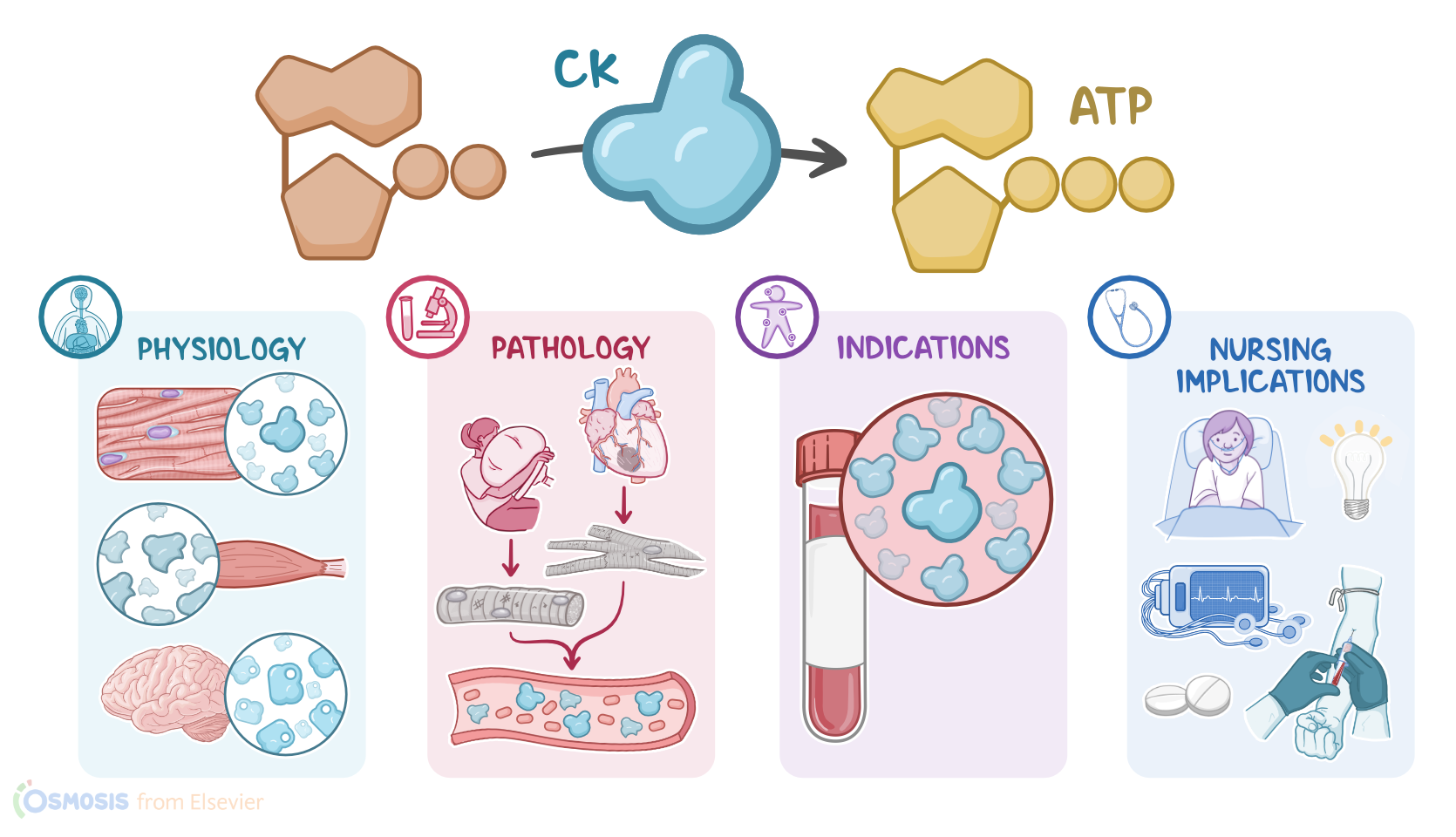
Okay, creatine kinase, or CK for short, also known as creatine phosphokinase or CPK, is an enzyme that helps create adenosine triphosphate, or ATP, which is an energy source, to supply to body tissues. Now, there are three types of CK: CK-MB, found mostly in heart muscle cells, or myocytes, with small amounts found in the skeletal muscle; CK-MM, found in skeletal muscle; and CK-BB, found in the brain.
Alright, let’s focus on the cardiac biomarker, CK-MB. There are certain conditions that can cause an increased CK-MB level, most commonly any condition that causes damage to cardiac or skeletal muscle cells.
Cardiac muscle cell damage can occur with acute coronary syndrome, which is any condition that decreases blood flow to the heart muscle, such as angina or myocardial infarctions, or MI for short. Decreased blood flow causes an imbalance between myocardial oxygen demand and supply from the coronary arteries, resulting in myocardial ischemia, a depletion of ATP, and a severe reduction in the ability of the heart to contract.
At the cellular level, damage to myocytes disrupts their membranes, causing the cellular contents, including CK-MB, to be released into the bloodstream. Now, when skeletal muscle cells are damaged, it can cause a condition called rhabdomyolysis, where the damaged muscle cells release their cellular contents into the bloodstream causing damage to the heart and kidneys.
Skeletal muscle can also be damaged by crushing trauma, which causes a traumatic form of rhabdomyolysis; and muscular dystrophy, a genetic condition causing progressive muscle weakness and loss of muscle mass.
Alright, so CK-MB is usually measured when there is a concern about cardiac ischemia or muscle damage.