Craniosynostosis: Nursing
Introduction0:00–0:18
Craniosynostosis is a congenital disorder characterized by the premature closure of a cranial sutures that may lead to deformity of the skull shape and inhibition of brain growth.Now, the skull consists of the paired frontal bones, the paired parietal bones, and the isolated occipital bone.
Physiology0:18–1:31
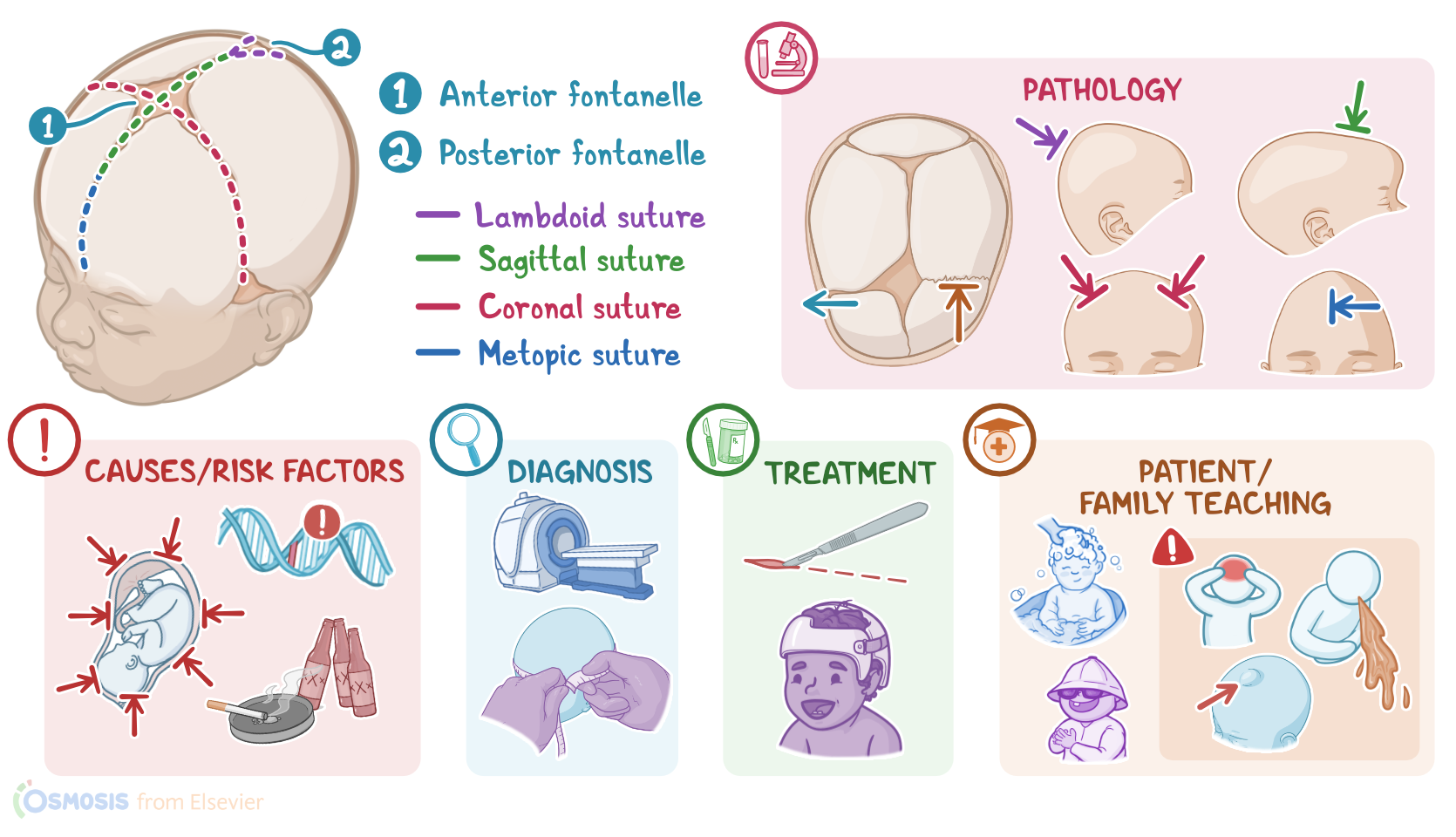
The spaces between the skull bones are called cranial sutures and there are four major sutures: metopic, coronal, sagittal, and lambdoid.
The sagittal, coronal, and metopic sutures meet at the anterior of the skull to form the anterior fontanelle, palpable just behind the forehead at the midline.
The posterior fontanelle is formed by the intersection of the sagittal and lambdoid sutures. At birth, the cranial bones and sutures are not well developed and are not fused to accommodate momentary skull distortion at birth and allow brain growth and development after birth.
At two months of age, the posterior fontanelle closes, followed by the closure of the anterior fontanelle at around 2 years old.
The metopic cranial suture also closes at two years of age and all other sutures close in adulthood upon completion of the craniofacial growth.Alright, now the exact causes of craniosynostosis are unknown, but it can be classified as non-syndromic, or isolated, which is the most common type, and syndromic, which is associated with other anomalies of the face, trunk, or extremities.Risk factors for craniosynostosis include, maternal use of tobacco, alcohol, or other substances during pregnancy, advanced maternal age, and family history of craniosynostosis.
Causes & risk factors1:31–2:20
Other causes include intrauterine constraint, where there’s pressure on the fetal skull during gestation, which can happen with maternal uterine malformations, multiple birth, or oligohydramnios, where there is not enough amniotic fluid to expand the uterine cavity.Okay, moving on to pathology, craniosynostosis occurs when there’s a premature fusion of one or more cranial sutures, which restricts the growth of the skull perpendicular to the affected suture.
Pathophysiology2:20–5:07
To accommodate the growth of the brain, the skull will grow parallel to the affected suture. The resulting skull deformity depends on the type of suture that is affected.
So, according to the affected suture, craniosynostosis can be classified into 5 types, including sagittal, metopic, unilateral coronal, bicoronal and lambdoid craniosynostosis.
Starting with sagittal craniosynostosis, also known as scaphocephaly, this is the most common form, and results from the premature fusion of the sagittal suture.
It’s characterized by a long and narrow growth of the head in the anteroposterior direction, and also a broad forehead and a prominent occiput.
Metopic craniosynostosis, also known as trigonocephaly, results from the premature fusion of the metopic suture, and is characterized by a triangular shaped forehead.
The eyebrows may appear pinched on either side, and the eyes may also appear close together. Down the forehead, a ridge can usually be felt.
Next, unilateral coronal craniosynostosis is known as anterior plagiocephaly, while lambdoid craniosynostosis is known as posterior plagiocephaly.
So, anterior plagiocephaly is caused by the early fusion of one side of the coronal suture, and is characterized by a flattened forehead and orbital rim on the affected side and a different shape of the eye on the affected side.
On the other hand, the posterior plagiocephaly is caused by the early fusion of the lambdoid suture and is characterized by a flattening of the back of the head.
Finally, bicoronal craniosynostosis, also known as brachycephaly, is caused by the early fusion of the coronal suture bilaterally.
It’s characterized by a wide shaped head with a very flat and tall forehead and the skull shortened in the anteroposterior direction.
Important complications associated with craniosynostosis include prolonged restriction of the cranial growth, which can result in increased intracranial pressure, or ICP, hydrocephalus, and inhibition of the brain growth.
Inhibition of brain growth can lead to impairment of the cognitive and neurodevelopment functions, and if the cranial nerves are involved, deficits in vision, hearing, and speech might occur.
In the terms of clinical manifestations, the first sign of craniosynostosis is an abnormally shaped skull which can vary depending on the affected suture.
Clinical manifestations5:07–5:31
Other signs may include an absence of soft spot on the baby’s skull, called a fontanelle; a raised firm edge where the sutures close; and delayed or absence of growth in the baby’s head size over time.
Diagnosis of craniosynostosis is mainly based on the client’s history and physical assessment. Imagistic studies such as X-ray, CT, or MRI might be also performed to further characterize the structural abnormalities.Treatment of craniosynostosis typically consists of a surgical procedure to correct the structural abnormalities, relieve pressure on the brain, and allow the brain to grow properly.
Diagnosis5:31–5:49
Treatment5:49–6:12
In cases of a very mild craniosynostosis, treatment may involve special medical helmets that help mold the baby’s skull into a more regular shape.
Alright, let’s look at the nursing care you’ll be providing for a child with craniosynostosis. Your priority nursing goals are to provide postoperative care, monitor for complications, and provide emotional support.
Management of care6:12–7:18
Begin by implementing routine postoperative interventions, and monitor them closely for complications related to the procedure.
Monitor for indications of increased ICP, and immediately report headache, altered mental status, visual disturbances, irritability, or nausea, and administer the prescribed treatments, as indicated.
Next, administer the prescribed prophylactic antibiotics, monitor the child’s temperature and report fever or other signs of infection like redness, swelling, or purulent exudate.
Finally, support the infant’s caregivers by providing emotional and psychological support. Encourage them to participate in care; and provide clear, concise answers to their questions and concerns.
Okay, let’s move on to client and family teaching. Begin by explaining that craniosynostosis occurs when the joints between the bones of their baby’s child's skull close too early.
General client & family teaching7:18–9:18
Teach them about each of their baby’s prescribed medications, and show them how to administer them exactly as directed. Review the plan of care and emphasize the importance of keeping all of their baby’s follow-up appointments for continued monitoring of their baby’s head circumference, growth, and development, as well as for ongoing physical, occupational, and speech therapy, as needed.
Then, remind them to get their baby fitted with a helmet to facilitate postoperative molding of their baby’s skull, and let them know that the helmet specialist will provide information on how to put the helmet on and take it off, and how long it should be worn each day.Then, teach the caregivers how to care for their baby at home.
Talk to them about the importance of providing adequate hydration and healthy nutrition to support recovery and growth. Instruct them to protect their baby from head injury by removing any low furniture with sharp edges.
Then, teach them how to care for their baby’s surgical site. Instruct them to keep it clean by gently washing the area with mild soap and water and patting it dry with a soft towel.
Also remind them to keep their baby’s head covered when outside, since the incision can become sunburned easily. Underscore the importance of contacting their healthcare provider immediately if they notice signs of infection, including increased redness and swelling, the presence of pus; if skin at the incision separates, or if their baby develops a fever.Also teach the caregivers how to monitor their baby’s head circumference, and to watch for signs of brain swelling.
Instruct them to seek emergency medical attention immediately if their baby develops headaches of vomiting; or has problems seeing or hearing.
Alright, as a quick recap…. Craniosynostosis refers to a congenital disorder characterized by premature closure of a cranial sutures, that can cause deformity of the skull shape and interference with normal growth of the brain.
Review9:18–10:28
Causes and risk factors include maternal uterine malformations, mechanical force on the skull during gestation, genetics, or teratogens.
Clinical manifestations include absence of soft spot on the baby’s skull, called a fontanelle, a raised firm edge where the sutures close, and delayed or no growth in the baby’s head size over time.
Diagnosis is established based on a thorough physical examination, as well as imaging studies such as X-rays, CT and MRI.
Depending on the severity of the condition, craniosynostosis can be treated surgically, or non-surgically by using specialized helmets that correct the condition as the infant child grows and develops.
Nursing goals include providing post-operative care, monitoring for complications, and providing emotional support. Client and family teaching is focused on the essentials of ongoing care, how to care for their baby at home, and when to seek medical care.
| CRANIOSYNOSTOSIS | ||
| KEY POINTS | NOTES | |
| DEFINITION |
| |
| PHYSIOLOGY |
| |
| CAUSES AND RISK FACTORS |
| |
| PATHOPHYSIOLOGY |
| |
| SIGNS AND SYMPTOMS |
| |
| DIAGNOSIS |
| |
| TREATMENT |
| |
| MANAGEMENT OF CARE |
| |
| PATIENT AND FAMILY TEACHING |
| |
No notes for this video yet
Try adding a note below