Electrocardiogram (ECG) - Normal sinus rhythm (NSR): Nursing
Introduction0:00–0:44
An electrocardiogram, commonly referred to as an ECG, is a routine noninvasive diagnostic test that uses electrodes placed on the skin to provide a graphic representation or picture of cardiac electrical activity.
The standard ECG, is also known as a 12-lead resting ECG because it has 12 leads or sets of electrodes. These include six limb leads, which are placed on the arms and legs, and six precordial leads, which are placed around the chest.
Keep in mind that a 12 lead ECG only uses 10 electrodes. That's because the chest electrodes record one lead each, while the limb detectors, together, actually record 6 leads.Now, let’s quickly review the electrophysiology of the heart.
Physiology0:44–2:50
The cardiac conduction system is made up of specialized myocardial cells that can create and send an electrical impulse, also called an action potential.
These cells have many special features, including automaticity, meaning they can generate an impulse by themselves; conductivity, meaning they can carry the impulse to other cells; and contractility, which is the ability to shorten the length of their fibers, causing a contraction.
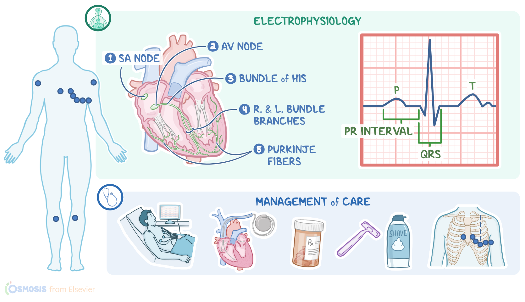
The way it goes is that the electric impulses begin in the sinoatrial or SA node, which is located at the junction of the superior vena cava and right atrium.
The SA node is considered the pacemaker of the heart and spontaneously and rhythmically produces impulses at 60 to 100 beats per minute.
The impulse then moves quickly through the atrial muscle causing depolarization. When the atrial muscle cells get depolarized, they contract, pushing blood from the atria into the ventricles.
Meanwhile, the impulse lands at the atrioventricular or AV node, which lies at the lower back section of the septum that separates the right and left atria.
Conduction velocity or the speed at which the impulse is propagated slows way down in the AV node. This allows time for the atria to contract while the ventricle fills.
From the AV node, the depolarization wave travels through the conducting system of the ventricles. First, it goes into the bundle of His, and then into the left and right bundle branches and into the Purkinje fibers.
The Purkinje fibers are the final bit of conductive tissue that spreads the depolarization wave to the rest of the heart.
The His-Purkinje system conducts the depolarization wave really quickly, and this is important because it makes the heart contract in a coordinated way.
This makes up the systole, where blood is ejected into the pulmonary and systemic circulations. Finally, the ventricles repolarize to prepare for the next cycle, which allows them to relax and fill with blood, called diastole.Now, the function of the heart’s conduction system is typically assessed using an ECG tracing, which shows how the depolarization wave moves during each heartbeat.
ECG reading2:50–4:29
So, in a typical waveform, there’s a P-wave, which represents the depolarization of the atria, a QRS complex, which represents depolarization of the ventricles; and a T-wave, which represents the repolarization of the ventricles.
Sometimes, immediately after the T-wave, there’s a U-wave, which represents late repolarization of the ventricles. In addition, there are certain intervals and segments.
So, there’s the PR interval, which spans from the beginning of the P-wave to the beginning of the QRS complex, and it represents the time it took for the impulse to travel from the SA node to the Purkinje fiber network.
And that’s actually the time from the beginning of atrial depolarization to the beginning of ventricular depolarization.
Within this time is the PR segment, the time between the end of the P-wave and the beginning of the QRS complex, which represents the time required for the impulse to travel through the AV node to the Purkinje fibers.
Then, there’s the QT interval, which spans from the beginning of the QRS complex to the end of the T-wave. It represents ventricular systole, which is the entire span from depolarization through repolarization.
Within the QT interval, there’s the ST segment which spans from the end of the QRS complex, called the J point, to the end of the T-wave and represents the time between ventricular depolarization and repolarization.Alright, so the findings of a normal ECG indicate a normal sinus rhythm and normal cardiac electrical activity.
Normal sinus rhythm4:29–6:22
The ECG paper has large squares with thick lines, and inside each are 25 smaller squares with thinner lines. Horizontally, each small square represents 0.04 seconds.
Vertically, each small square represents 0.1 millivolts.There are eight main things to look for on an ECG to ensure everything is normal.
First, the heart rate can be calculated by counting the number of QRS complexes in six seconds and multiplying that number by 10.
Secondly, the heart rhythm regularity can be assessed by checking the distance between the peaks of two consecutive P waves and two consecutive R waves.
If the ratio of the P-P and R-R intervals is 1, then the heart rhythm is regular. Thirdly, the P wave should be checked to ensure it is present, regular in timing and shape, and that there is one for each QRS complex.
Fourth, the PR interval is checked to make sure it’s consistently between 0.12 to 0.20 seconds, which is three to five little boxes.
Fifth, the QRS duration should be consistently less than 0.1 seconds, which is two and a half little boxes. Sixth, the ST segment should not be elevated or depressed more than 1 small box.
Seventh, the T wave shape should be asymmetric and height should be less than 5 millimeters in precordial leads and less than 10 millimeters in limb leads.
And finally, the QT interval should be less than or equal to half of a cardiac cycle, which can be calculated by measuring the distance between two successive R waves.Alright, let’s take a look at the nursing care you’ll be providing for a client who is receiving an electrocardiogram test with a 12 lead ECG.
Management of care6:22–8:33
Start by explaining that an ECG test analyzes the electrical activity of the heart to diagnose various heart conditions.
Remind your client that this routine procedure is painless, and there is no electric shock involvement during the recording.
To prepare them for the procedure, be sure to ensure their safety and ability to follow directions. Then, ask your client if they have any implanted pacemaker devices or if they are taking medications that can alter heart function, such as tricyclic antidepressants, beta blockers, calcium channel blockers, or diuretics.
Next, inspect the skin, making sure it is clean and dry, and shave or clip any excess chest hair, which would interfere with the test.
Next, attach the electrodes to their specific location on your client’s skin. So, first, place the RA lead on the right arm or shoulder, and place the LA lead on the left arm or shoulder; then, the RL goes on the right leg, while the LL is placed on the left leg.
Now for the precordial leads, Lead V1 is placed on the right sternal border at the fourth intercostal space, while Lead V2 goes on the left sternal border at the fourth intercostal space; Lead V4 is placed at the midclavicular line at the fifth intercostal space and then Lead V3 goes midway between V4 and V2; Lead V5 goes on the anterior axillary line at the fifth intercostal space; and lastly, Lead V6 goes on the midaxillary line, fifth intercostal space at the same level as V4.Be sure all the connections are secure to prevent any artifact that would cause an inaccurate ECG test.
Then, assist your client into a semi-reclined posture with legs uncrossed, and ensure that they can remain still and breathe normally during the test.
When the test is completed remember to remove all the electrodes off your client’s skin, and ensure your client’s safety.
Alright, as a quick recap. The 12 lead ECG is a routine, noninvasive test that uses electrodes placed on the skin to provide a graphic representation of the electrical activity of the heart.
Review8:33–8:58
This can be used to diagnose various cardiac conditions. The proper placement of the electrodes on the skin and elimination of artifacts during the recording are the most important in obtaining an accurate ECG for the healthcare provider to interpret
| ELECTROCARDIOGRAM (ECG) - NORMAL SINUS RHYTHM (NSR) | ||
| KEY POINTS | NOTES | |
| DEFINITION |
| |
| PHYSIOLOGY |
| |
| ECG READING |
| |
| NORMAL SINUS RHYTHM |
| |
| MANAGEMENT OF CARE |
| |
No notes for this video yet
Try adding a note below