Fetal circulation: Nursing
Introduction0:00–0:33
Fetal circulation involves the delivery of oxygen and nutrients from the placenta to the fetus and the transport of waste products from the fetus to the placenta, in order for them to be eventually eliminated by the mother’s body.
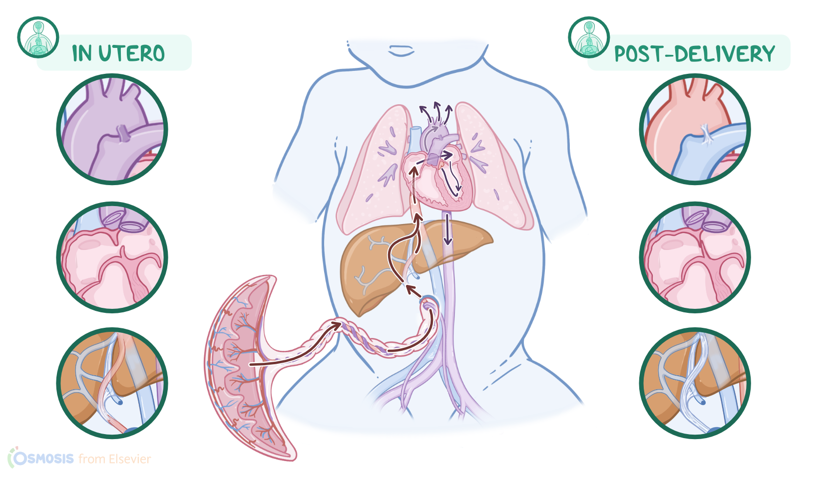
Fetal circulation has some unique features because the fetal lungs are not involved in gas exchange and there are three fetal shunts, which redirect the blood to ensure the highest oxygenated blood reaches the heart and brain, while redirecting blood away from the nonfunctional fetal lungs.
Now let’s first review the physiology of the umbilical cord that connects the fetus to the placenta, which takes on the role of exchanging oxygen, carbon dioxide, nutrients, and wastes.
Physiology0:33–3:18
The cord houses two umbilical arteries and one umbilical vein. The umbilical vein provides the path for oxygenated blood to flow from the placenta to the fetus, while the umbilical arteries carry deoxygenated blood back to the placenta.
Now, oxygenated blood first flows through the umbilical vein and to the fetal liver, and here, the circulation divides as it meets the first fetal shunt called the ductus venosus.
At this point, most of the blood passes through the ductus venosus and flows directly into the inferior vena cava. Meanwhile, the remaining blood perfuses the liver and then meets up with the rest of the blood in the inferior vena cava.
Together, this blood flows into the right atrium of the fetal heart. In the fetal heart, the pressure on the right side is higher than on the left side of the heart.
This is because the fetal lungs are filled with fluid and the arteries are tightly constricted, so the pressure in the fetal lungs is high, leading to increased pressure in the right side of the heart.This pressure difference allows a majority of the oxygenated blood in the right atrium to pass through the second shunt, called the foramen ovale, which is an opening between the two atria.
This shunt allows most of the blood to bypass the lungs and flow directly into the left atrium. From here, the blood flows into the left ventricle, which pumps it through the aorta, to supply oxygenated blood to perfuse the brain, the heart, and then the rest of the fetus.
At the same time, the right ventricle pumps blood into the pulmonary trunk. Again, since the lungs are collapsed, only a small portion of oxygenated blood enters the lungs.
The remaining blood flows through the third shunt, called the ductus arteriosus, into the descending aorta and eventually to the umbilical arteries that lead to the placenta.After birth, when the baby takes its first breath, their lungs expand and pulmonary blood flow increases.
This makes pulmonary vessels expand, and decreases pulmonary pressure. In turn, this decreases the pressure in the right side of the heart and the change in pressures causes the foramen ovale to close.
As the baby continues to breathe, arterial oxygen levels increase, causing the ductus arteriosus to constrict. When the umbilical cord is cut, the placental circulation is separated from the fetal circulation, causing the ductus venosus to constrict.
The process of closing these three shunts is gradual and may take up to 48 hours to fully close.Alright, as a quick recap… The adaptations specific to fetal circulation include three shunts, or passages, called the ductus venosus, the foramen ovale, and the ductus arteriosus.Oxygenated blood flows from the placenta through the umbilical vein to reach the fetal liver.
Review3:18–4:18
Here, most of the oxygenated blood passes through the ductus venosus directly into the inferior vena cava, while the rest perfuses the liver and meets with the rest of the blood in the inferior vena cava.
This blood is then carried into the right atrium, and passes through the foramen ovale into the left atrium. The oxygenated blood flows into the left ventricle, which pumps it through the aorta and first to the brain and then the rest of the body.
Finally, since the lungs are collapsed in utero, they only get a small part of oxygenated blood; and the rest flows through the ductus arteriosus and into the descending aorta.
After delivery, these shunts usually close on their own in up to 48 hours.
| FETAL CIRCULATION | ||
| KEY POINTS | NOTES | |
| DEFINITION |
| |
| PHYSIOLOGY |
| |
No notes for this video yet
Try adding a note below