Hip fractures: Nursing
Introduction0:00–0:30
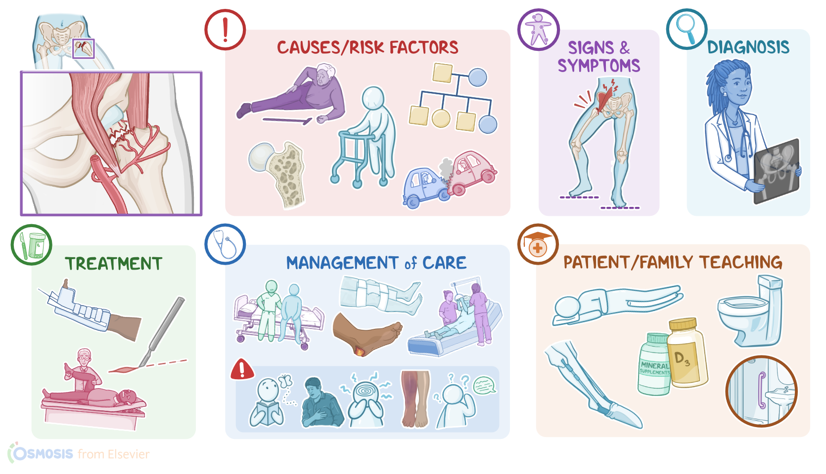
Hip fractures are fractures that occur in the upper third of the thigh bone, called the femur. These fractures can be grouped into two types: intracapsular fractures, which are fractures that happen within the capsule of the hip joint; and extracapsular fractures, which are fractures that happen outside the capsule.
One of the most common sites for a hip fracture is at the femoral neck and this is a type of intracapsular fracture. Now, the hip joint, also called the acetabulofemoral joint, is a ball and socket joint formed between the acetabulum of the hip bone and the head of the femur.
Physiology0:30–1:02
The articulating bones are covered by hyaline cartilage and a joint capsule, which is reinforced on the outside by strong ligaments.
This makes the joint strong and stable, allowing it to connect the free bones of the lower limb to the pelvic bones. The hip joint allows multiaxial movements, but is also important for weight-bearing and providing stability.
Now, intracapsular fractures are caused by trauma and osteoporosis. Low bone mass due to osteoporosis causes the bones to become more fragile, making them more prone to breaking.
Causes & risk factors1:02–1:44
Because of this, intracapsular fractures are also called fragility fractures, because they typically occur after minimal trauma, like falling from a chair.
On the other hand, extracapsular fractures are typically caused by severe direct trauma, like a car crash. Risk factors for hip fractures include old age, which can lead to falls, being assigned female at birth, and having a family history of osteoporosis.Now, the main blood supply for the femoral head comes from the lateral femoral circumflex artery that travels along the femoral head.
Pathophysiology1:44–2:19
This artery is often compromised during a hip fracture which can cause complications like avascular necrosis of the femoral head.
Furthermore, reduced blood flow means slower healing which leads to longer periods of immobility and hospitalization. This increases the risk of developing thrombotic complications, like deep vein thrombosis and pulmonary emboli as well as other complications like infections.
Clients with a hip fracture typically experience severe pain and tenderness around the affected area which makes it difficult to bear weight on the affected leg.
Clinical manifestations2:19–2:39
Sometimes, abduction and external rotation of the hip joint, muscle spasm, and shortening of the affected limb might be seen.
The diagnosis of hip fractures starts with the client's history and physical assessment, followed by imaging tests. These include anteroposterior and lateral plain X-rays.
Diagnosis2:39–2:53
Treatment2:53–3:46
The treatment of hip fractures includes acute management followed by rehabilitation. Acute management includes controlling pain using a regional nerve block, immobilization of the hip joint.
Buck’s traction, which keeps the leg in an extended position, is sometimes used. Often, surgical repair and stabilization of the displaced bones is required.
Anticoagulants may also be administered to prevent thrombosis and avascular necrosis. Now, rehabilitation can take place in a rehabilitation unit, a skilled nursing facility, or at home, and consists of physical and occupational therapy.
Physical therapy involves early, frequent exercises that restore strength and tone of hip muscles, whereas occupational therapy helps with activities of daily living.
Alright, let’s look at the nursing care you’ll be providing for a client with a hip fracture. Your priority goals of care are to prepare your client for surgery, monitor for postoperative complications, and promote mobility.Begin preparing your client for surgery by immobilizing the affected extremity and helping them into a comfortable position.
Management and care3:46–7:54
Assess their pain level and manage their pain by assisting with the insertion of a catheter nerve block and administering a bolus injection of a local anesthetic like bupivacaine, as prescribed.
Establish IV access and administer the prescribed prophylactic antibiotic. After surgical repair, provide standard post-operative care and monitor them closely for complications related to the procedure.
Institute fall precautions, maintain bed rest with the affected extremity immobilized and abducted in the prescribed position, and ensure that the head of the bed is not elevated more than 45 degrees to prevent excessive hip flexion.
Also continue IV fluids and antibiotics, and manage their pain, as needed. Routinely assess their vital signs and surgical dressing, reinforcing it as needed.
Report to the healthcare provider if you notice signs of infection, excessive discharge or bleeding from the wound; as well as the development of fever, hypotension, or tachycardia.
Remember to be vigilant about the prevention of pressure injuries by ensuring your client is placed on a pressure-redistributing surface, monitoring their pressure points, and assisting with position changes at least every 2 hours.
Also, encourage them to use the overhead trapeze bar to shift position as needed. Now, if your client is elderly, remember they are at increased risk of delirium during hospitalization.
Be sure to monitor them closely for signs of delirium such as a decreased ability to focus, disorientation, or disturbances in their understanding of language, and compare their current mental status with their baseline.
If you suspect delirium, provide reassurance, and report your findings to the healthcare provider right away.Alright, you’ll also need to keep a close eye on your client’s affected extremity by regularly performing a full neurovascular assessment.
Immediately report to the healthcare provider if your client develops cool, pale skin, diminished distal pulses, prolonged capillary refill, or impaired sensation or motor function.
Also administer the prescribed venous thromboembolism prophylaxis, monitor for signs of deep vein thrombosis or pulmonary embolism, and immediately report if your client experiences leg pain or swelling; as well as shortness of breath, cough, or chest pain.Also, keep in mind that early mobilization can prevent postoperative complications and speed your client’s recovery.
So you’ll be working with the physical therapist to assist your client to sit at the edge of the bed, stand, transfer to a chair, and walk with the use of an assistive device such as a walker.
Remember to assess your client’s pain level and administer the prescribed analgesics as needed prior to activity. During therapy sessions, help them to protect their operative site by avoiding flexing their hip more than 90 degrees while sitting, or crossing their surgical leg across their body’s midline.
Report to the healthcare provider if your client has signs of dislocation, including sudden, severe pain, limb shortening, and external rotation of the limb.
Finally, collaborate with your unit’s case manager to coordinate your client’s care transition to a rehabilitation facility or home, and arrange for continued physical therapy, home health care, adaptive equipment, and transportation to appointments, as needed.
Okay, moving on to client and family teaching. Begin by explaining where their fracture occurred, how it was repaired, and the plan of care for their recovery and rehabilitation.
General client and family teaching7:54–11:21
Teach them about each of the medications they will be taking at home, including how and when to take them, and side effects they should be aware of.
If they’re prescribed an anticoagulant, instruct them to watch for signs of bleeding, and tell them to contact their healthcare provider right away if they notice easy bruising or bleeding from their nose or gums; likewise, instruct them to contact their healthcare provider if they notice signs of a clot, such as pain, redness, or swelling in their calf, thigh, or groin; or if their foot gets cold or changes color.Next, remind them how participating in their physical therapy appointments and doing exercises at home, will help build strength and balance so they can move normally again.
Then, review how to keep their hip in proper alignment by avoiding actions such as crossing their legs or ankles when they sit or are lying down; bending at their hips more than 90 degrees, which is like the angle in the letter "L"; or lifting their knee higher than their hip.
Also remind them to keep their knees and toes pointed forward when they sit, walk, or stand. Then, explain how keeping a pillow between their knees when they’re in bed will help prevent their surgical leg from crossing over the midline of their body; and show them how using a raised toilet seat extender can help them avoid excessive bending at the hip when using the bathroom.
Then, ensure they know how to use adaptive devices to support their independence, like a sock aid. Lastly, instruct them to immediately notify their healthcare provider for any signs of hip dislocation such as sudden hip pain; numbness of the affected leg; difficulty bearing weight on the affected leg; if the leg appears shorter; or if it looks like it is turned inwards or outwards.
Remind them to monitor their surgical incision site, and to let their healthcare provider know if they have signs or symptoms of an infection, including increased pain, swelling, or warmth; if they notice pus is draining from the incision; or if they develop a fever.
And lastly, instruct them to seek medical attention right away for signs of avascular necrosis, such as a gradual onset of throbbing hip pain that may radiate to the groin, buttocks, or even down to their knee.Finally, review some simple lifestyle modifications they can do to decrease the risk of future fractures.
Also teach them about maintaining strong bones by engaging in regular weight-bearing exercises as tolerated, including sources of protein, calcium, and vitamin D in their diet, and to continue to take their prescribed calcium and vitamin D supplements, and bisphosphonates.
Be sure to provide them with a list of recommended foods and sample menus to help them adhere to a bone-healthy diet. Lastly, teach them about the importance of limiting alcohol; and if they need help with smoking cessation, provide them with counseling and resources for ongoing support.Alright, as a quick recap….
Review11:21–12:46
Hip fractures occur in the upper third of the thigh bone. Hip fractures can be intracapsular and extracapsular, and one of the most common sites is a femoral neck fracture, which is a type of intracapsular fracture.
Intracapsular fractures typically occur secondary to minimal trauma and osteoporosis, while extracapsular neck fractures are typically only caused by severe direct trauma.
Clients with a hip fracture typically experience severe pain and tenderness around the affected area which makes it difficult to bear weight on the affected leg.
Client history, physical assessment, and diagnostic imaging are used for diagnosing hip fractures. Acute management of hip fractures includes controlling pain using a regional nerve block, immobilization of the joint, and Buck’s traction can also be done to keep the leg in an extended position.
Surgical repair and stabilization are often required. Rehabilitation after surgery includes physical therapy and occupational therapy.
Goals of nursing care are to prepare your client for surgery, monitor for postoperative complications, and promote mobility.
Client and family education includes positioning precautions, lifestyle modifications for maintaining bone health, preventing future hip fractures, learning about postoperative complications and when
| HIP FACTURES | ||
| KEY POINTS | NOTES | |
| DEFINITION |
| |
| PHYSIOLOGY |
| |
| CAUSES AND RISK FACTORS |
| |
| PATHOPHYSIOLOGY |
| |
| SIGNS AND SYMPTOMS |
| |
| DIAGNOSIS |
| |
| TREATMENT |
| |
| MANAGEMENT OF CARE |
| |
| PATIENT AND FAMILY TEACHING |
| |
No notes for this video yet
Try adding a note below