Nutrition - Enteral: Nursing skills

1,788views

Nutrition - Enteral: Nursing skills

Week 10 modules

Week 10 modules

Diabetes mellitus: Pathology review
Diabetes mellitus
Diabetes mellitus (DM): Nursing process (ADPIE)
Hyperosmolar hyperglycemic state (HHS): Nursing process (ADPIE)
Diabetic ketoacidosis (DKA): Nursing process (ADPIE)
Case study - Diabetic ketoacidosis (DKA): Nursing
Medication administration - Insulin: Nursing pharmacology
Insulin: Nursing pharmacology
Anatomy of the abdominal viscera: Esophagus and stomach
Anatomy of the abdominal viscera: Large intestine
Anatomy of the abdominal viscera: Small intestine
Renal system anatomy and physiology
Anatomy of the urinary organs of the pelvis
Malnutrition: Nursing
Diarrhea: Nursing
Assessment - Nutrition: Nursing
Urinary retention: Nursing
Case study - Constipation: Nursing
Obesity: Nursing
Glucagon
Glycogen metabolism
Peripheral arterial disease (PAD): Nursing process (ADPIE)
Peripheral venous disease (PVD): Nursing process (ADPIE)
Non-insulin injectable antidiabetic drugs - GLP-1 agonists and amylinomimetics: Nursing pharmacology
Oral antidiabetic medications - Alpha-glucosidase inhibitors: Nursing pharmacology
Nutrition - Enteral: Nursing skills
Oral antidiabetic medications - DPP-4 inhibitors: Nursing pharmacology
Oral antidiabetic medications - Sulfonylureas and meglitinides: Nursing pharmacology
Oral antidiabetic medications - Sodium-glucose co-transporter-2 (SGLT-2) inhibitors: Nursing pharmacology
Carbohydrates and sugars
Gastrointestinal system anatomy and physiology
Proteins
Fats and lipids
Vitamins and minerals
Urinary incontinence - Stress: Nursing process (ADPIE)
Hygiene - Ostomy care: Nursing skills
Routine ostomy care: Clinical skills notes
Urinary tract infections (UTIs): Nursing process (ADPIE)
GI/GU: Assisting with bowel elimination
Approach to hypoglycemia: Clinical sciences
Diabetic ketoacidosis: Clinical sciences
Hyperosmolar hyperglycemic state: Clinical sciences
Complications of Diabetes
Video Case Study - Bowel Elimination
Approach to diarrhea (chronic): Clinical sciences
Nursing Care for Enteral Nutrition
Hydration
Malnutrition
Obesity and Health Risks
Obesity and metabolic syndrome: Clinical sciences
Protein-calorie malnutrition: Clinical sciences
Bladder and bowel training: Clinical skills notes
GI/GU: Bladder and bowel training
Lower urinary tract infection

Notes

NUTRITION - ENTERAL

KEY POINTS
NOTES
DEFINITION
  • Delivers fluids and nutrients into GI tract 
  • Used for patients with chewing or swallowing issues 
  • Common conditions  
    • Head or neck trauma 
    • Surgery
    • Coma 
    • Dementia
    • Nervous system disorders 
    • Tumors of head neck or esophagus 
  • Types of feeding tubes 
    • Nasogastric (NG) tube 
      • Goes through nose into stomach 
      • Used for short term feeding 
    • Naso-intestinal (NI) tube 
      • Goes through nose into small intestine 
      • Also used for short term feeding 
    • Gastrostomy (G) tube 
      • Inserted through abdominal wall into stomach 
      • Used for long term feeding 
      • Percutaneous endoscopic gastrostomy (PEG) tube 
        • Inserted into stomach using endoscopic guidance 
    • Jejunostomy (J) tube 
      • Inserted into small intestine (jejunum) 
      • Used for long term feeding 
  • Duration of use 
    • NG and NI tubes: short term 
      • Few days - 6 weeks 
    • G tubes PEG tubes J tubes: long term 
      • > 6 weeks

FEEDING SCHEDULES
  • Confirm tube placement 
    • Verified by X ray
  • Start enteral nutrition 
    • Begin feeding slowly 
    • Watch for nausea, pain, or diarrhea 
    • Increase rate if feeding is tolerated 
  • Types of nutritional formulas 
    • Chosen based on patient needs and condition 
  • Feeding administration methods 
    • Infusion pump 
      • Continuous feeding 
        • Runs throughout day 
        • Provides steady nutrient delivery 
      • Intermittent feeding 
        • Scheduled at intervals during day 
        • Mimics normal meal patterns

ADMINISTERING ENTERAL FEEDINGS
  • Before starting 
    • Review order and verify correct formula 
    • Identify patient
    • Explain procedure 
    • Answer questions 
    • Perform hand hygiene
    • Apply clean gloves 
  • Verify tube placement 
    • Feeding tube tip may shift locations 
    • Draw up 30 mL of air into 60 mL syringe 
    • Instill air into tube and aspirate 5–10 mL fluid 
    • Observe fluid appearance 
      • Gastric: clear pale yellow or green 
      • With formula: looks like curdled milk 
      • Intestinal: golden yellow to greenish brown 
    • Test pH with indicator strip 
      • Gastric: pH 0–4 if fasting 
      • Continuous feeding: pH 5–6 
      • pH > 6 may indicate wrong placement 
    • Hold feeding if placement is uncertain 
  • Prepare formula and equipment 
    • Open system 
      • Use cans of formula and administration bag 
      • Check expiration date and shake cans 
      • Clean tops and pour into bag 
      • Clamp tubing before filling 
    • Closed system 
      • Use prefilled container and administration set 
      • Check expiration date and shake container 
      • Remove cover and spike with tubing 
  • Administer feeding 
    • Raise head of bed 30–45 degrees 
    • Hang formula 12 inches above patient’s head 
    • Prime tubing and insert into feeding pump 
    • Cleanse tube end and flush with 30 mL water 
    • Connect administration set to feeding tube 
    • Program pump and start feeding 
    • Check for kinks or blockages if alarm sounds 
    • After feeding, flush tube with 30 mL water 
    • Cap tube and keep patient upright for 1 hour 
  • Alternative feeding methods 
  • Gravity feeding with roller clamp 
    • Slower and may be better tolerated 
  • Bolus feeding with syringe 
    • Use 60 mL syringe over 10 minutes 
    • Pour formula into graduated cylinder 
    • Deliver by gravity through syringe 
  • Check gastric residual volume (GRV) 
    • Connect syringe and aspirate stomach contents 
    • Note and document residual amount 
    • GRV > 250 mL may indicate intolerance 
    • Return contents to prevent malnutrition

CLINICAL IMPLICATIONS
  • NG and NI tubes
    • Can irritate nasal mucosa 
    • Use approved device to secure tube 
    • Provide skin care as needed 
    • Insertion site care 
      • Keep site clean and dry 
      • Watch for redness tenderness or pain 
      • Apply protective skin barrier if needed 
    • Maintain tube patency 
      • Flush with water per facility policy 
      • Helps prevent clogging 
  • Report and respond to problems
    • Insertion site issues 
      • Fluid leaking around G tube J tube or PEG tube
      • Inability to confirm tube placement 
    • Feeding intolerance signs 
      • Gastric residual volume too high 
      • Abdominal pain, nausea, or distension
    • Aspiration signs 
      • Coughing
      • Choking
      • Gagging
      • Breathing difficulty 
  • Preventing error
    • Never use IV lines for enteral feeding 
      • Can cause severe harm or death 
    • Use ENFit connectors only 
      • Designed to prevent misconnections 
      • Incompatible with IV or other non enteral devices
    • Label and trace tubing 
    • Use labels “Tube Feed Only” 
    • Trace tubing to confirm enteral route

Transcript

Watch video only

Enteral nutrition provides fluids and nutrients directly into the GI tract through a feeding tube. As a healthcare professional, you’ll be responsible for caring for patients receiving enteral nutrition.

Now, enteral nutrition might be especially helpful for patients who have difficulty chewing or swallowing as a result of head or neck trauma; surgery; coma; dementia; nervous system disorders; or tumors of the head, neck, or esophagus.

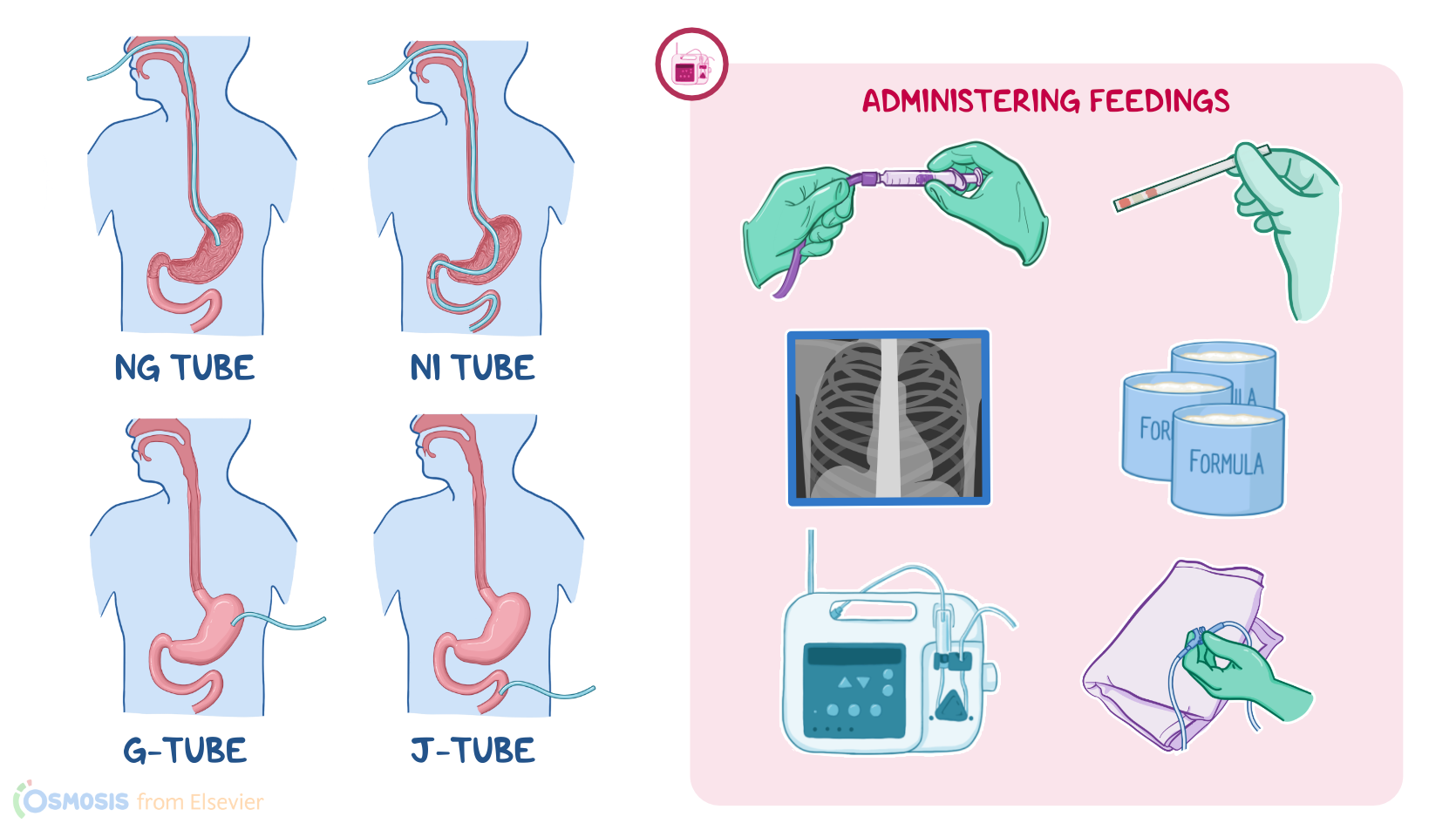
Now, there are various ways a feeding tube can go into the GI tract. The most common one is a nasogastric, or NG, tube that goes through the nose and then down into the stomach. A naso-intestinal, or NI tube, also goes through the nose, but it ends in the small intestines.

Tubes can also be inserted through a surgical opening, or stoma, in the abdominal wall and into the stomach, known as a gastrostomy tube, or G-tube. A common type of gastrostomy tube is a percutaneous endoscopic gastrostomy tube, or PEG tube for short, that’s inserted directly into the stomach percutaneously with endoscopic guidance. Finally, tubes can also be inserted into a part of the small intestine called the jejunum, known as a jejunostomy tube, or J-tube.

J-tubes, G-tubes, and PEG tubes are usually inserted if long-term tube feedings are anticipated and are typically used for more than six weeks; whereas NG and NI tubes are mostly preferred when tube feedings are needed for a short period of time, typically a few days or up to six weeks.

Once the feeding tube is inserted, its placement is confirmed by X-ray. After that, your patient can start receiving enteral nutrition through the tube. There are different types of nutritional formulas that can be ordered depending on your patient’s needs. When you first start a feeding, you’ll usually begin feeding slowly while you watch for nausea, abdominal pain, or diarrhea that could indicate feeding intolerance. If the initial rate and amount of feeding is tolerated, the feedings can be increased to the desired amount.

Now, enteral nutrition is commonly administered using an infusion pump. In this case, administration schedules can be either continuous or intermittent. A continuous feeding is when a patient has feedings running through their feeding tube throughout the day. On the flip side, if feedings are scheduled at intervals over the course of the day, this is called an intermittent feeding.

Before starting a feeding, first review the patient’s chart, collect your supplies, and make sure you have the correct formula. Identify your patient, inform them about the procedure, and answer any questions related to the procedure. Then, perform hand hygiene and apply clean gloves.

Now, keep in mind that sometimes the tip of the feeding tube can move to a different location, like from the stomach to the intestines or esophagus, or from the intestines to the stomach. So, you’ll need to verify the tube’s position before starting the feeding by checking the appearance and pH of fluid aspirated through the tube.

To do this, draw up 30 mL of air into a 60 mL enteral syringe, connect the syringe to the end of your patient’s tube and gently instill the air. Then, slowly retract the plunger and aspirate 5 to 10 mL of fluid, transfer it to a medicine cup, and observe the fluid’s appearance. Gastric fluid can range in color from clear and colorless or pale yellow or green if your patient has been fasting at least four hours. If there’s still formula in the stomach, the fluid will look like curdled milk. Intestinal fluid is often stained by bile, so color can range from golden yellow to greenish brown.

Next, test the pH by applying a few drops of fluid onto a pH indicator strip, or you can dip the strip into the medicine cup. Use a color chart to determine the pH. For gastric fluid, a pH of 0 to 4 is normal if your patient has been fasting for at least four hours; a pH 5 to 6 may occur if they’re receiving continuous feedings; and a pH more than 6 means the tube is in the esophagus, lung, or small intestines. If the pH does not match the desired location of the tube, the feeding should be held until the tube is replaced or placement is confirmed with imaging.

Once tube placement is confirmed, prepare to administer the feeding. For feedings that will run through an enteral feeding pump, you’ll use either an open or closed system. If you’re using an open system, you’ll need cans of the prescribed formula, an administration bag, and an administration set.

First, check the expiration date, then shake the cans, and cleanse the tops before opening them. Attach the tubing to the bag, clamp it to prevent the formula from running through the tube, and pour the formula into the bag.

Sources

  1. "Clinical nursing skills & techniques (11th ed.). ISBN 978-0-443-10718-4 " Elsevier (2025)
  2. "Fundamentals of nursing (12th ed.). " Elsevier (2026)
  3. "Alexander’s Nursing Practice: Hospital and Home. 5th edition. ISBN: 978-0-7020-6230-8 " Elsevier (2019)
  4. "Clinical Nursing Skills and Techniques - E-Book. 10th ed. ISBN 978032379653 " Elsevier Health Sciences (2021)
  5. "Fundamentals of Nursing. 11th edition. ISBN: 978-0-323-81034-0 " Elsevier (2022)
  6. "A guide to enteral nutrition in intensive care units: 10 expert tips for the daily practice. 25(1):424. Published 2021 Dec 14. " Crit Care (2021)
  7. "Krause and Mahan’s Food and the Nutrition Care Process. 16th edition. ISBN: 978-0-323-81025-8 " Elsevier Canada (2022)
  8. "Critical Care Nursing: Diagnosis and Management. 9th edition. ISBN: 978-0-323-64295-8 " Elsevier (2021)