Physical assessment - Eyes: Nursing

Last updated: July 26, 2022

Physical assessment - Eyes: Nursing

Watch later

Watch later

Cholinomimetics: Direct agonists
Immunosuppressants for autoimmune diseases: Nursing pharmacology
Osteoporosis: Nursing
Osteomyelitis: Nursing
Paget disease of bone: Nursing
Cerebral palsy: Nursing
Down syndrome (Trisomy 21)
Encephalitis: Nursing
Headaches: Pathology review
Meningitis: Nursing process (ADPIE)
Seizure disorder: Nursing process (ADPIE)
Stroke: Nursing process (ADPIE)
Erysipelas and cellulitis: Nursing
Antibiotics - Penicillins: Nursing pharmacology
Antifungals - Topical: Nursing pharmacology
Cutaneous fungal infections: Nursing
Antibiotics - Cephalosporins: Nursing pharmacology
Antibiotics - Macrolides: Nursing pharmacology
Antivirals for influenza: Nursing pharmacology
Antivirals for herpesviruses: Nursing pharmacology
Antivirals for hepatitis B and C: Nursing pharmacology
Antibiotics - Fluoroquinolones: Nursing pharmacology
Antibiotics - Tetracyclines and glycylcyclines: Nursing pharmacology
Antidepressants - Tricyclic antidepressants (TCAs) and monoamine oxidase inhibitors (MAOIs): Nursing pharmacology
Glaucoma: Nursing process (ADPIE)
Antidepressants - SSRIs and SNRIs: Nursing pharmacology
Gout: Nursing process (ADPIE)
Antiretrovirals for HIV/AIDS - CCR5 antagonists, fusion inhibitors, and attachment inhibitors: Nursing pharmacology
Antiretrovirals for HIV/AIDS - Integrase strand transfer inhibitors: Nursing pharmacology
Antiretrovirals for HIV/AIDS - Protease inhibitors: Nursing pharmacology
Antiretrovirals for HIV/AIDS - NRTIs and NNRTIs: Nursing pharmacology
Cataracts: Nursing
Detached retina: Nursing
Macular degeneration: Nursing
Meniere disease: Nursing
Growth and development - Infant: Nursing
Otitis media: Nursing
Eye anesthetics: Nursing pharmacology
Mydriatics and cycloplegics: Nursing pharmacology
Ophthalmic anti-inflammatories and anti-infectives: Nursing pharmacology
Diabetes mellitus (DM): Nursing process (ADPIE)
Anatomy and physiology of the eye
Physical assessment - Eyes: Nursing
Conjunctivitis: Nursing process (ADPIE)
Eye conditions: Inflammation, infections and trauma: Pathology review
Anatomy of the eye
Pediatric psychosocial needs during illness and hospitalization: Nursing
Hearing impairment and otosclerosis: Nursing
Growth and development theories: Nursing
Growth and development - Preschool-age: Nursing
Growth and development - Toddler: Nursing
Growth and development - School-age: Nursing
Growth and development - Adolescent: Nursing
Antibiotics - Carbapenems and monobactams: Nursing pharmacology
Antibiotics - Trimethoprim and sulfonamides: Nursing pharmacology
Antibiotics - Beta lactam and beta lactamase inhibitor combinations: Nursing pharmacology
Antibiotics - Aminoglycosides: Nursing pharmacology
Medications for respiratory syncytial virus (RSV): Nursing pharmacology
Mycobacterium tuberculosis (Tuberculosis)
Hypersensitivity reactions - Type II: Nursing
Lyme disease: Nursing process (ADPIE)
Systemic lupus erythematosus (SLE): Nursing
Preoperative care: Nursing
Postoperative care: Nursing
Chest tube care: Nursing
Analgesics for obstetrics: Nursing pharmacology
Analgesics: Nursing pharmacology
Acne: Nursing
Burn injury: Nursing
Impetigo: Nursing
Insect stings and bites: Nursing
Psoriasis: Nursing
Herpes zoster: Nursing
Urticaria: Nursing
Pressure injury: Nursing process (ADPIE)
Cholinergic therapy (GU): Nursing pharmacology
Physical assessment - Skin, hair, and nails: Nursing
Acetylcholinesterase inhibitors for myasthenia gravis: Nursing pharmacology
Antiepileptics: Nursing pharmacology
Narcolepsy (NORD)
Autism spectrum disorder (ASD): Nursing
Insomnia
Hemorrhagic stroke - Intracranial hemorrhage (ICH) and subarachnoid hemorrhage (SAH): Nursing
Body fluid compartments
Anemia - Macrocytic: Nursing
Anemia - Iron-deficiency: Nursing
Vitamin D
Vitamin B12 deficiency
Role of Vitamin K in coagulation
Folate (Vitamin B9) deficiency
Coronary artery disease (CAD) and angina pectoris: Nursing process (ADPIE)
Hypertension: Nursing process (ADPIE)
Hypersensitivity reactions - Type I: Nursing
Hypersensitivity reactions - Type III: Nursing
Hypersensitivity reactions - Type IV: Nursing
Immune response - Adaptive: Nursing
Immune response - Innate: Nursing
Human immunodeficiency virus (HIV) and acquired immunodeficiency syndrome (AIDS): Nursing
Antiglaucoma medications: Nursing pharmacology
Oral antidiabetic medications - Sodium-glucose co-transporter-2 (SGLT-2) inhibitors: Nursing pharmacology
Insulin: Nursing pharmacology
Non-insulin injectable antidiabetic drugs - GLP-1 agonists and amylinomimetics: Nursing pharmacology
Oral antidiabetic medications - Biguanides and thiazolidinediones: Nursing pharmacology
Oral antidiabetic medications - Sulfonylureas and meglitinides: Nursing pharmacology
Oral antidiabetic medications - DPP-4 inhibitors: Nursing pharmacology
Oral antidiabetic medications - Alpha-glucosidase inhibitors: Nursing pharmacology
Antihistamines: Nursing pharmacology
Antacids: Nursing pharmacology
Antiemetics: Nursing pharmacology
Gastric mucosal protective agents: Nursing pharmacology
Antidiarrheals: Nursing pharmacology
Laxatives: Nursing pharmacology
Antispasmodics (GI): Nursing pharmacology
Chronic obstructive pulmonary disease (COPD): Nursing process (ADPIE)
Physiology of pain: Nursing

Notes

PHYSICAL ASSESSMENT - EYES

KEY POINTS
NOTES
DEFINITION
  • Completed as part of comprehensive or focused assessment 
  • Gives information about vision and eye health
  • Identifies ocular problems early

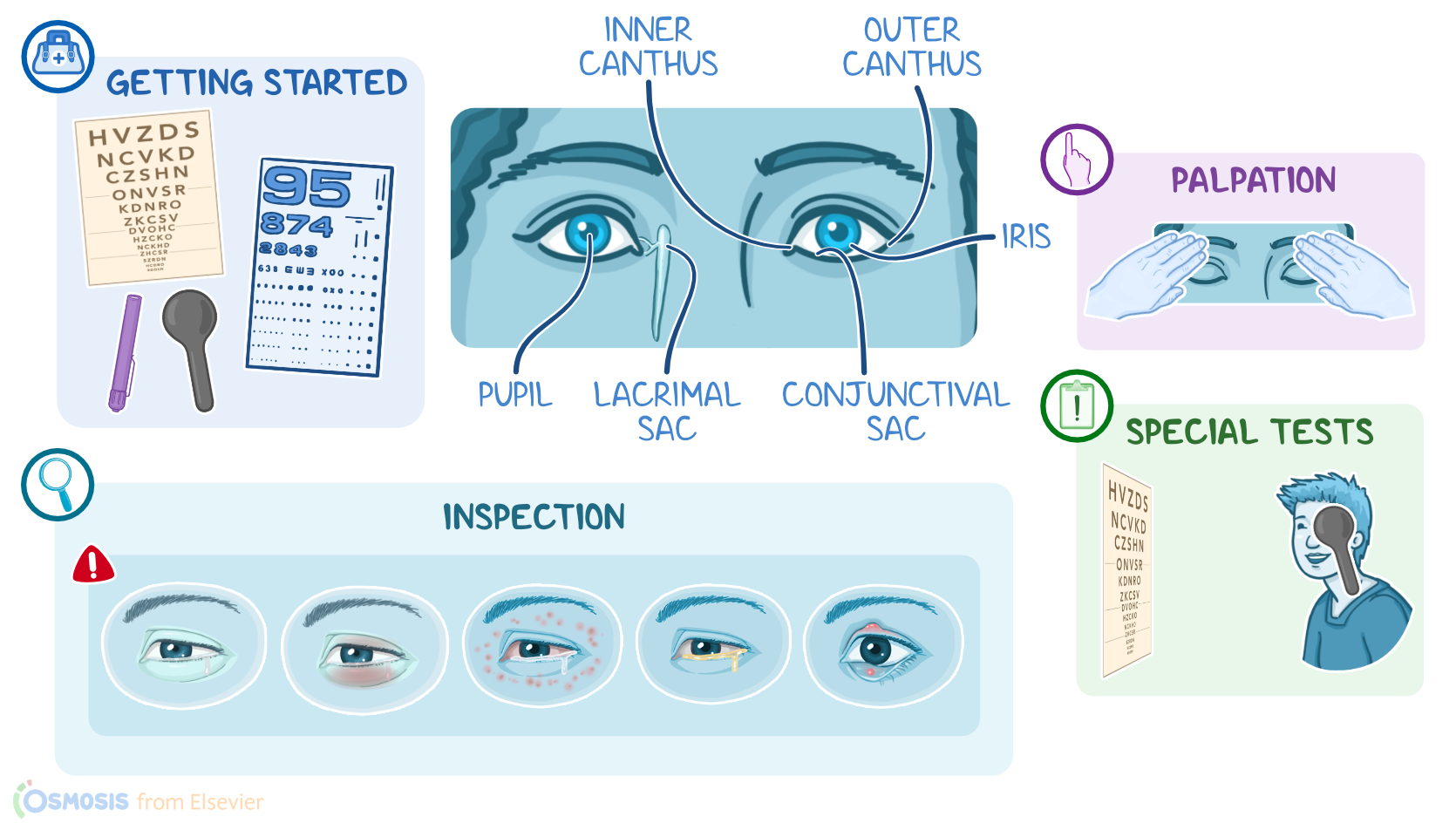
GETTING STARTED
  • Supplies
    • Snellen/Sloan chart
    • Rosenbaum/Jaeger near vision card
    • Penlight
    • Eye cover 
  • Preparation
    • Adequate light
    • Ensure patient comfort
    • Explain procedure
    • Answer questions
    • Obtain verbal consent 
    • Hand hygiene 
    • Collect supplies
    • Provide privacy

ANATOMICAL LANDMARKS
  • Upper eyelids
  • Lower eyelids
  • Eyebrows
  • Inner canthus
  • Outer canthus
  • Pupil
  • Lacrimal sac
  • Conjunctival sac
  • Iris

METHODS OF ASSESSMENT
  • Inspection
  • Palpation
  • Special tests

INSPECTION
  • External eyes
    • Begin at eyebrows and move downward 
    • Eyelashes and eyebrows should be even 
    • Eyelids should open and close fully 
    • Upper eyelids should cover eyes equally 
    • Abnormal findings 
      • Drooping upper eyelid (ptosis)
        • May result from neuromuscular weakness 
        • Can be linked to cranial nerve three damage 
  • Surrounding structures 
    • Should be free of edema or puffiness 
    • No lesions drainage or nodules present 
    • Xanthelasma  
      • May indicate abnormal lipid metabolism 
  • Sclera and conjunctiva 
    • Sclera should normally appear white 
      • Yellow sclera may suggest liver disease 
    • Conjunctiva should be clear and moist 
      • Redness may indicate conjunctivitis 
  • Iris and lens 
    • Iris should be round and evenly colored 
    • Shine penlight to view lens clarity 
      • Cloudy lens may indicate cataracts 
  • Pupils 
    • Pupils should be round and equal bilaterally 
    • Dim lights to allow pupil dilation 
    • Shine penlight into one eye 
      • Observe for direct constriction 
      • Check for consensual constriction in other eye 
    • Pupil abnormalities 
      • Miosis is abnormal pupil constriction 
        • Often caused by opioid use 
      • Anisocoria is unequal pupil size 
        • Can be normal or disease related 
  • Extraocular muscle strength 
    • Tests cranial nerves III, IV, & VII
      • Ask patient to follow finger with eyes 
      • Move through 6 cardinal gaze fields 
      • Pause at each extreme for one to two seconds 
      • Watch for smooth symmetrical eye movement 
      • Nystagmus  
        • May be normal or indicate muscle weakness 
  • Cover-uncover test 
    • Patient looks at fixed point ahead 
    • Cover one eye and observe other 
      • Uncovered eye should stay focused 
    • Remove cover and observe again 
      • Gaze should remain steady 
    • Strabismus is eye deviation inward 
      • Caused by weak extraocular muscles

PALPATION
  • Palpate 
    • Eyelids
    • Surrounding structures 
  • Feel for nodules, warmth, or discomfort
  • Eyes should be soft, gently pushed on w/o discomfort
  • Resistant to palpation
    • Possible glaucoma or tumor

SPECIAL TESTS
  • Visual acuity  
    • Tests function of cranial nerve II
    • Use Snellen or Sloan chart for distance vision 
    • Patient stands 20 ft from chart 
    • Cover one eye and read letters 
    • Repeat with other eye 
    • Record vision as a fraction 
      • Top number is patient distance from chart 
      • Bottom number is normal reading distance 
    • Larger denominator means worse visual acuity 
    • Vision worse than 20/200 is blindness 
  • Near vision testing 
    • Use Rosenbaum or Jaeger handheld card 
    • Hold card 14 inches from eyes 
    • Patient reads letters or numbers aloud 
    • Stop when letters become too small

NURSING IMPLICATIONS
  • Assess
  • Interpret
  • Document
  • Report abnormal findings to HCP
  • Monitor patient progress and changes from baseline

Transcript

Watch video only

Assessment of the eyes should be completed as part of a comprehensive client assessment or as part of a focused exam when a client is experiencing ocular issues, such as eye pain or blurred vision. This assessment gives the nurse information about vision and general eye health while helping to identify ocular problems at an early stage, such as glaucoma or cataracts. Assessment of the eyes includes several tests which will examine the eye itself as well as screen for ocular diseases or systemic diseases that manifest through the eye, like diabetes or liver disease. Let’s review the process of completing an eye assessment.

Okay, the supplies you’ll need for the eye assessment include a Snellen or Sloan chart, a Rosenbaum or Jaeger near vision card, a penlight, and an eye cover. You should prepare for the eye exam by ensuring you have adequate light, and that your client is comfortable in either a standing or sitting position.

Before getting started, explain the procedure to your client and be sure to answer any questions they might have before obtaining verbal consent. Then, perform hand hygiene and collect your supplies.

Now, locating the anatomical landmarks of the eyes and surrounding tissue will help guide your assessment. These landmarks include the upper eyelids and lower eyelids, eyebrows, the inner canthus and the outer canthus, pupil, lacrimal sac, conjunctival sac, and iris.

Alright, the methods of ocular assessment include inspection and palpation as well as a series of visual tests.

First, you should inspect the external eyes and the surrounding structures, starting at the eyebrows and moving downward. Eyelashes and eyebrows should be evenly distributed. Eyelids should be able to close and open all the way, and upper eyelids should extend equally over both eyes.

If an upper eyelid droops and partially covers the eye, ptosis is present, which may be due to neuromuscular weakness from conditions such as myasthenia gravis or damage to cranial nerve III.

The surrounding structures of the eyes should be without edema, puffiness, lesions, drainage or nodules.

Raised, misshaped, yellow lesions on the surrounding eye tissue, called xanthelasma, may indicate abnormal lipid metabolism.

You’ll also inspect the sclera and conjunctiva for redness and vascularity. The sclera should normally be white but it can appear yellow in clients with liver disease. The conjunctiva should be translucent, but can become pinkish-red in clients with conjunctivitis.

The iris should be round with an even distribution of color. When shining your penlight into the eye, the lens should be transparent; but if it appears cloudy, this can indicate cataracts.