Physical assessment - Peripheral vascular system: Nursing

1,292views

Physical assessment - Peripheral vascular system: Nursing

NRS 243

NRS 243

Respiratory system anatomy and physiology
Pneumonia
Chest tube care: Nursing
Physical assessment - Thorax and lungs: Nursing
Pulmonary hypertension
Pulmonary embolism
Cor pulmonale
Pulmonary edema: Nursing
Bronchodilators: Nursing pharmacology
Cardiac preload
Cardiac afterload
Cardiac contractility
Cardiac work
Geriatric considerations - Cardiac: Nursing
Cardiac biomarkers - Troponin: Nursing
Mitral valve disease
Hypertension: Nursing process (ADPIE)
Blood pressure: Clinical skills notes
Cardiovascular system anatomy and physiology
Normal heart sounds
ECG basics
ECG rate and rhythm
ECG intervals
Action potentials in pacemaker cells
Action potentials in myocytes
Excitability and refractory periods
Cardiac excitation-contraction coupling
Cardiac conduction system
Stroke volume, ejection fraction, and cardiac output
Blood pressure, blood flow, and resistance
Compliance of blood vessels
Resistance to blood flow
Renin-angiotensin-aldosterone system
Baroreceptors
Chemoreceptors
Abnormal heart sounds
Anatomy of the coronary circulation
Heart failure: Pathology review
Aortic valve disease
Valvular heart disease: Nursing
Peripheral arterial disease (PAD): Nursing process (ADPIE)
Peripheral venous disease (PVD): Nursing process (ADPIE)
Physical assessment - Peripheral vascular system: Nursing
Buerger disease: Nursing
Shock - Obstructive: Nursing
Shock - Anaphylactic: Nursing
Shock - Neurogenic: Nursing
Shock - Hypovolemic: Nursing
Shock - Cardiogenic: Nursing
Shock - Septic: Nursing
Arrhythmias - Asystole: Nursing
Arrhythmias - Atrial fibrillation (Afib): Nursing
Arrhythmias - Atrial flutter (Aflutter): Nursing
Arrhythmias - Heart blocks: Nursing
Arrhythmias - Premature atrial contractions (PACs): Nursing
Arrhythmias - Premature ventricular contractions (PVCs): Nursing
Arrhythmias - Sinus tachycardia and sinus bradycardia: Nursing
Arrhythmias - Supraventricular tachycardia (SVT): Nursing
Arrhythmias - Ventricular fibrillation (Vfib): Nursing
Arrhythmias - Ventricular tachycardia (Vtach): Nursing
Arterial embolism: Nursing
Electrocardiogram (ECG) - Normal sinus rhythm (NSR): Nursing
Cardiomyopathy: Nursing
Congenital heart defects - Acyanotic: Nursing
Congenital heart defects - Cyanotic: Nursing
Endocarditis: Nursing
Heart defects that decrease pulmonary blood flow - Nursing considerations & client education: Nursing
Kawasaki disease: Nursing
Myocarditis: Nursing
Pericarditis: Nursing
Aortic aneurysm: Nursing process (ADPIE)
Coronary artery disease (CAD) and angina pectoris: Nursing process (ADPIE)
Left-sided heart failure: Nursing process (ADPIE)
Myocardial infarction (MI): Nursing process (ADPIE)
Pericardial effusion and cardiac tamponade: Nursing process (ADPIE)
Rheumatic heart disease: Nursing process (ADPIE)
Alpha-1 adrenergic blockers: Nursing pharmacology
Alpha-2 adrenergic agonists: Nursing pharmacology
Angiotensin II receptor blockers (ARBs): Nursing pharmacology
Angiotensin-converting enzyme (ACE) inhibitors: Nursing pharmacology
Antiarrhythmics: Nursing pharmacology
Antihyperlipidemics - Fibrates: Nursing pharmacology
Antihyperlipidemics - Miscellaneous: Nursing pharmacology
Antihyperlipidemics - Statins: Nursing pharmacology
Beta-adrenergic blockers: Nursing pharmacology
Calcium-channel blockers: Nursing pharmacology
Cardiac glycosides: Nursing pharmacology
Direct-acting vasodilators: Nursing pharmacology
Nitrates: Nursing pharmacology
Sympathomimetic medications: Nursing pharmacology
Pharyngitis: Nursing
Foreign body aspiration and upper airway obstruction: Nursing process (ADPIE)
Pneumothorax and hemothorax: Nursing
Chronic obstructive pulmonary disease (COPD): Nursing process (ADPIE)
Chronic disease: Nursing
Chronic bronchitis
Acute respiratory distress syndrome (ARDS): Nursing
Disaster management: Nursing
Cardiac cycle
Cardiac biomarkers - Creatine kinase (CK): Nursing
Laryngeal cancer: Nursing
Tracheostomy suctioning: Clinical skills notes
Care of an intubated client: Nursing skills
Anatomy of the larynx and trachea
Corticosteroids - Inhaled: Nursing pharmacology
Microcirculation and Starling forces
Pressures in the cardiovascular system
Tracheostomy: Nursing

Notes

PHYSICAL ASSESSMENT - PERIPHERAL VASCULAR SYSTEM

KEY POINTS
NOTES
DEFINITION
  • Completed as part of a comprehensive or focused assessment 

GETTING STARTED
  • Supplies 
    • Stethoscope with diaphragm and bell
    • Skin marker
    • Doppler ultrasound device
    • Drapes
    • Light source
  • Preparation 
    • Adequate light 
    • Ensure patient comfort 
    • Explain procedure 
    • Answer questions 
    • Provide privacy 
    • Obtain verbal consent 
    • Hand hygiene 
    • Collect supplies 

ANATOMICAL LANDMARKS
  • Carotid pulse 
    • Found on neck behind sternocleidomastoid muscle 
    • Just below angle of jaw 
  • Brachial pulse 
    • Located in cubital fossa center 
    • Medial to biceps tendon 
  • Radial pulse 
    • Found at wrist lateral forearm 
    • Just below base of thumb 
  • Femoral pulse 
    • Below inguinal ligament 
    • Between pubic and hip bones 
  • Popliteal pulse 
    • Located behind the knees 
  • Dorsalis pedis pulse 
    • Found on top of the foot 
  • Posterior tibial pulse 
    • Located behind medial malleolus

METHODS OF ASSESSMENT
  • Inspection
  • Palpation
  • Auscultation

INSPECTION
  • Look for symmetry between right and left sides 
  • Asymmetry may indicate abnormal findings 
  • Skin 
    • Light skin pallor appears pale 
    • Dark skin pallor appears ashen or gray 
    • Brown skin pallor may show yellow undertones 
    • Check palmar surfaces for paleness in dark skin
    • Ruddy color may suggest vascular disorder 
    • Redness may indicate localized infection 
  • Signs of vascular dysfunction 
    • Varicose veins are enlarged and twisted 
    • Venous ulcers appear near medial malleolus 
    • Arterial ulcers often found on toes 
    • Look for jugular venous distention (JVD)
      • May suggest fluid volume overload

PALPATION
  • Begin palpation with temperature check 
    • Use back of hands for assessment 
    • Skin should feel warm and consistent 
    • Cool areas may suggest poor perfusion 
    • Warm areas may indicate possible infection 
  • Peripheral pulses 
    • Use pads of two fingers 
    • Assess each pulse point bilaterally 
    • Palpate simultaneously when possible 
    • Do not palpate carotids at same time 
    • Avoid excess pressure on carotid pulse 
    • Prevent vagal stimulation or fainting 
    • Evaluate pulse rhythm and strength 
    • Grade pulse intensity 
      • 0 pulse is absent 
      • 1 + is weak and thready 
      • 2 + is normal strength 
      • 3 + is strong and full 
      • 4 + is bounding pulse 
      • Use doppler if pulse is hard to find 
        • Helpful in poor perfusion or obesity 
        • Listen for whooshing sound 
        • Mark pulse location with skin marker 
        • Grade as 0 if pulse not found 
  • Capillary refill 
    • Press nail bed until it blanches 
    • Release and count color return time 
    • Normal refill is < 2 seconds 
    • Delayed refill may suggest poor circulation 
  • Edema
    • Press over tibia or top of foot 
    • Look for indentation after pressure 
    • Grade edema
      • 0 + means no edema 
      • 1 + is 2mm rebounds immediately 
      • 2 + is 3 - 4mm rebounds in 15 seconds 
      • 3 + is 5 - 6mm rebounds in 60 seconds 
      • 4 + is 8mm rebounds in 2 - 3 minutes 

AUSCULTATION
  • Use stethoscope bell
  • Listen over carotid and femoral arteries
  • Bruit is abnormal finding

NURSING IMPLICATIONS
  • Assess 
  • Interpret 
  • Document 
  • Report abnormal findings to HCP 
  • Monitor patient progress and changes from baseline

Transcript

Watch video only

Assessment of the peripheral vascular system should be completed as part of a comprehensive client assessment, or as part of a focused exam if the client is experiencing issues that might be related to the function of the peripheral vascular system, like arterial or venous ulcers. Let’s review the process of completing an assessment of the peripheral vascular system.

Okay, the supplies you’ll need for your assessment include a stethoscope with a diaphragm and bell, a skin marker, a doppler ultrasound device, drapes, and a good source of light.

Then, prepare for the exam by ensuring your client is in a comfortable position, that your hands and stethoscope are warm, and that the temperature in the room is comfortable. Provide privacy by closing the door and curtains, properly draping your client, and only exposing areas of their body as needed to perform your examination.

Before getting started, explain the procedure to your client and be sure to answer any questions they might have before obtaining verbal consent. Then, perform hand hygiene and collect your supplies.

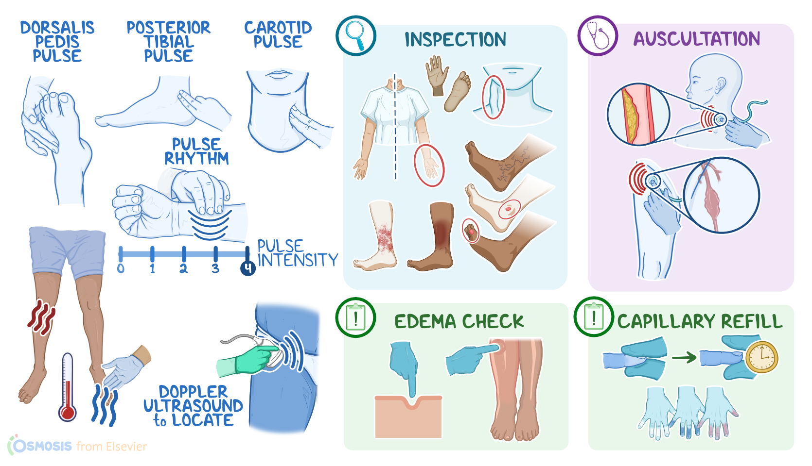
Now, locating the anatomical landmarks of the peripheral vascular system will help guide your assessment. Peripheral pulses that can be palpated include the carotid pulse, located on the neck behind the sternocleidomastoid muscle, or scm, just below the angle of the jaw; the brachial pulse, located in the center of the cubital fossa, medially to the biceps tendon; the radial pulse, found in the wrist along the lateral aspect of the forearm, just below the base of the thumb; the femoral pulse, located below the inguinal ligament, between the pubic and hip bones; the popliteal pulse, located behind the knees; the dorsalis pedis pulse, found on the dorsal aspect of the foot; and the posterior tibial pulse, located just behind the medial malleolus.

Alright, methods of assessment for the peripheral vascular system include inspection, palpation, and auscultation.

Let’s start with inspection. During your assessment, remember to look for symmetry between the right and left sides, since an abnormal finding might be present in one side and not the other.

Look for signs of adequate perfusion by observing the color of your client's extremities. Pallor, which may indicate poor arterial perfusion, will present as a pale color in clients with light skin; in clients with dark skin, pallor may present as a more ashen or gray color; while in clients with brown skin, pallor may have yellowish undertones. Another method to assess for pallor in darker skin tones is to inspect the palmar surfaces which might appear more pale.

A dark, ruddy discoloration might indicate a vascular disorder like venous insufficiency; and an erythematous or red appearance could indicate a localized infection.

Next, look for obvious signs of peripheral vascular dysfunction like varicose veins, which are enlarged, tortuous veins most often found in the lower extremities; venous ulcerations, which typically present at the medial malleolus; or arterial ulcers, that are commonly found on the toes.

Finally, inspect the jugular veins on the neck for any signs of jugular venous distention, or JVD, as this could indicate fluid volume overload, associated with problems like heart or liver failure.

Next, move on to palpation. Assess the temperature of the upper and lower extremities, by using the back of your hands. Normally, the temperature of the skin should be warm and relatively consistent in the upper and lower extremities.

If there are localized areas where the skin is cool to the touch, this can be an indication of impaired perfusion. On the other hand, if the skin feels unusually warm, an infection might be present.