Pleural effusion: Nursing

1,527views

Pleural effusion: Nursing

Acute Final

Acute Final

Endocrine system anatomy and physiology
Antepartum assessment - Fetus: Nursing
Assessment of gestational age: Nursing
Fetal circulation: Nursing
Fetal development: Nursing
Group B streptococcus (GBS) infection in pregnancy: Nursing
Hepatitis B virus (HBV) infection in pregnancy: Nursing
Hyperemesis gravidarum: Nursing
Large for gestational age (LGA) infant: Nursing
Preeclampsia and eclampsia: Nursing
Prenatal screening: Nursing
Placenta previa: Nursing process (ADPIE)
Placental abruption: Nursing process (ADPIE)
Birth-related procedures: Nursing
Cesarean birth: Nursing
Intrapartum assessment - Fetal heart rate patterns: Nursing
Intrapartum assessment - Uterine activity: Nursing
Premature rupture of membranes (PROM): Nursing
Shoulder dystocia: Nursing
Prolapsed umbilical cord: Nursing process (ADPIE)
Stages of labor: Nursing
Assessment - Postpartum: Nursing
Perinatal depression: Nursing
Physiology of lactation: Nursing
Postpartum infections: Nursing
Postpartum hemorrhage: Nursing
Biliary atresia: Nursing
Cleft lip and palate: Nursing
Congenital diaphragmatic hernia: Nursing
Congenital heart defects - Acyanotic: Nursing
Congenital heart defects - Cyanotic: Nursing
Esophageal atresia and tracheoesophageal fistula: Nursing
Craniosynostosis: Nursing
Hemolytic disease of the fetus and newborn: Nursing
Hyperbilirubinemia: Nursing process (ADPIE)
Infant of a diabetic mother (IDM): Nursing
Meconium aspiration syndrome: Nursing
Neonatal respiratory distress syndrome (NRDS): Nursing
Neonatal sepsis: Nursing
Neural tube defects: Nursing
Newborn adaptation to extrauterine life: Nursing
Persistent pulmonary hypertension of the newborn (PPHN): Nursing
Physical assessment - Neonate: Nursing
Small for gestational age (SGA) infant: Nursing
Postterm infant: Nursing
Thermoregulation - Neonate: Nursing
Arterial blood gas (ABG) - Overview: Nursing
Arterial blood gas (ABG) - Metabolic acidosis: Nursing
Arterial blood gas (ABG) - Metabolic alkalosis: Nursing
Arterial blood gas (ABG) - Respiratory acidosis: Nursing
Arterial blood gas (ABG) - Respiratory alkalosis: Nursing
Adrenal insufficiency (Addison disease): Nursing
Anemia - Iron-deficiency: Nursing
Anemia - Aplastic: Nursing
Anemia - Macrocytic: Nursing
Case study - Hypothyroidism: Nursing
Case study - Iron-deficiency anemia: Nursing
Case study - Sickle cell anemia: Nursing
Complete blood count (CBC) - Hemoglobin and hematocrit: Nursing
Complete blood count (CBC) - Red blood cells (RBC): Nursing
Complete blood count (CBC) - Platelets: Nursing
Complete metabolic panel (CMP) - Blood urea nitrogen (BUN) and creatinine (Cr): Nursing
Complete metabolic panel (CMP) - Estimated glomerular filtration rate (eGFR): Nursing
Complete metabolic panel (CMP) - Liver function tests (LFT): Nursing
Cushing syndrome and Cushing disease: Nursing
Hematopoietic growth factors: Nursing pharmacology
Hyperparathyroidism: Nursing
Hyperthyroidism: Nursing process (ADPIE)
Hypoparathyroidism: Nursing
Hyperpituitarism: Nursing
Hypopituitarism: Nursing
Hypothyroidism: Nursing process (ADPIE)
Medications affecting the parathyroid glands: Nursing pharmacology
Medications for growth hormone disorders: Nursing pharmacology
Medications for thyroid disorders: Nursing pharmacology
Neutropenia: Nursing
Polycythemia: Nursing
Thrombocytopenia: Nursing
Acute kidney injury (AKI): Nursing process (ADPIE)
Benign prostatic hyperplasia (BPH): Nursing process (ADPIE)
Case study - Cholecystitis: Nursing
Case study - Cirrhosis: Nursing
Case study - Chronic kidney disease (CKD): Nursing
Case study - Benign prostatic hyperplasia (BPH): Nursing
Case study - Gastroesophageal reflux disease (GERD): Nursing
Case study - Pediatric appendicitis: Nursing
Case study - Pyelonephritis: Nursing
Cholecystitis: Nursing
Cholelithiasis: Nursing
Chronic kidney disease (CKD): Nursing
Cirrhosis: Nursing process (ADPIE)
Diverticular disease: Nursing
Gastroesophageal reflux disease (GERD): Nursing process (ADPIE)
Hemolytic uremic syndrome: Nursing
Hirschsprung disease: Nursing
Intestinal obstruction: Nursing
Irritable bowel syndrome (IBS): Nursing
Nephrotic syndrome: Nursing
Pyloric stenosis: Nursing process (ADPIE)
Renal and urinary calculi: Nursing
Urinary incontinence - Stress: Nursing process (ADPIE)
Diabetes insipidus: Nursing process (ADPIE)
Dialysis care: Nursing
Case study - Diabetic ketoacidosis (DKA): Nursing
Case study - Pediatric diabetes mellitus type 1: Nursing
Diabetes mellitus (DM): Nursing process (ADPIE)
Hyperosmolar hyperglycemic state (HHS): Nursing process (ADPIE)
Diabetic ketoacidosis (DKA): Nursing process (ADPIE)
Case study - Epilepsy: Nursing
Case study - Head injury: Nursing
Epidural and subdural hematoma: Nursing
Case study - Stroke: Nursing
Hemorrhagic stroke - Intracranial hemorrhage (ICH) and subarachnoid hemorrhage (SAH): Nursing
Increased intracranial pressure (ICP): Nursing
Hydrocephalus: Nursing process (ADPIE)
Intracranial aneurysm: Nursing
Seizure disorder: Nursing process (ADPIE)
Stroke: Nursing process (ADPIE)
Jaundice: Nursing
Nutrition - Enteral: Nursing skills
Nutrition - Newborn: Nursing
Nutrition - Parenteral: Nursing skills
Phenylketonuria (PKU): Nursing
Arterial embolism: Nursing
Disseminated intravascular coagulation (DIC): Nursing
Hemophilia: Nursing process (ADPIE)
Acute respiratory distress syndrome (ARDS): Nursing
Asthma: Nursing process (ADPIE)
Atelectasis: Nursing
Bacterial pneumonia: Nursing process (ADPIE)
Bronchiolitis and respiratory syncytial virus (RSV): Nursing process (ADPIE)
Case study - Acute respiratory distress syndrome (ARDS): Nursing
Care of an intubated client: Nursing skills
Case study - Chronic obstructive pulmonary disease (COPD): Nursing
Case study - Impaired gas exchange: Nursing
Case study - Pediatric asthma: Nursing
Chest tube care: Nursing
Chronic obstructive pulmonary disease (COPD): Nursing process (ADPIE)
Cystic fibrosis: Nursing
Epiglottitis: Nursing process (ADPIE)
Flail chest: Nursing
Intraoperative care: Nursing
Pleural effusion: Nursing
Pneumothorax and hemothorax: Nursing
Pulmonary edema: Nursing
Smoke inhalation injury: Nursing process (ADPIE)
Tracheostomy: Nursing
Venous thromboembolism (VTE): Nursing process (ADPIE)
Arrhythmias - Asystole: Nursing
Arrhythmias - Atrial flutter (Aflutter): Nursing
Arrhythmias - Premature atrial contractions (PACs): Nursing
Arrhythmias - Heart blocks: Nursing
Arrhythmias - Atrial fibrillation (Afib): Nursing
Arrhythmias - Premature ventricular contractions (PVCs): Nursing
Arrhythmias - Sinus tachycardia and sinus bradycardia: Nursing
Arrhythmias - Supraventricular tachycardia (SVT): Nursing
Arrhythmias - Ventricular fibrillation (Vfib): Nursing
Arrhythmias - Ventricular tachycardia (Vtach): Nursing
Cardiac biomarkers - Troponin: Nursing
Case study - Acute coronary syndrome (ACS): Nursing
Case study - Atrial fibrillation (Afib): Nursing
Case study - Heart failure with reduced ejection fraction (HFrEF): Nursing
Case study - Deep vein thrombosis (DVT): Nursing
Case study - Hypertension: Nursing
Case study - Hypovolemic shock: Nursing
Coronary artery disease (CAD) and angina pectoris: Nursing process (ADPIE)
Electrocardiogram (ECG) - Normal sinus rhythm (NSR): Nursing
Heart defects that decrease pulmonary blood flow - Nursing considerations & client education: Nursing
Hypertension: Nursing process (ADPIE)
Left-sided heart failure: Nursing process (ADPIE)
Myocardial infarction (MI): Nursing process (ADPIE)
Pericardial effusion and cardiac tamponade: Nursing process (ADPIE)
Peripheral arterial disease (PAD): Nursing process (ADPIE)
Rheumatic heart disease: Nursing process (ADPIE)
Shock - Cardiogenic: Nursing
Shock - Neurogenic: Nursing
Shock - Obstructive: Nursing
Shock - Septic: Nursing
Sickle cell disease: Nursing process (ADPIE)
Valvular heart disease: Nursing

Notes

PLEURAL EFFUSION

KEY POINTS
NOTES
DEFINITION
  • Excess fluid accumulated between visceral layer and parietal layer of pleura

PHYSIOLOGY
  • Pleural space fluid
    • 20-25 mL fluid for lubrication
    • Forms as filtrate from pleural blood vessels
    • Drains into lymphatic vessels

CAUSES AND RISK FACTORS
  • Causes
    • Increased production or impaired drainage
      • Transudate
      • Exudate
  • Risk factors
    • Underlying heart, lung, kidney, or liver conditions
    • Exposure to tobacco smoke, alcohol
    • Use of certain medications

PATHOPHYSIOLOGY
  • Excess fluid builds in pleural space 
  • Pressure placed on lungs and surrounding structures
  • Impaired ventilation and oxygenation 
  • Impaired drainage of pleural fluid allows bacteria to grow

SIGNS AND SYMPTOMS
  • Asymptomatic
  • Symptomatic
    • Dyspnea
    • Dry cough
    • Sharp pain with inspiration
    • Reduced or absent breath sounds
    • Dullness to percussion

DIAGNOSIS
  • History
  • Physical assessment
  • X-ray
  • CT
  • Thoracentesis

TREATMENT
  • Therapeutic thoracentesis
  • Chest tube 
  • Surgery
  • Address underlying cause

MANAGEMENT OF CARE
  • Goals of care
    • Improve respiration and oxygenation
    • Monitor for complications 
  • Elevate head of bed
  • Encourage coughing, deep breathing, and use of incentive spirometer
  • Monitor respiratory status
  • Auscultate lungs
  • Assess skin and nails
  • Apply oxygen as needed
  • Notify HCP
    • Increased work of breathing
    • Cyanosis
    • Oxygen saturation below baseline
  • Thoracentesis
    • Reinforce procedure information
    • Confirm informed consent
    • Evaluate puncture site
    • Assess pain
    • Administer medications as prescribed
    • Monitor vital signs, respiratory efforts, and oxygen saturation
    • Monitor for complications
    • Notify HCP
      • Manifestations of pneumothorax

PATIENT AND FAMILY TEACHING
  • Explain condition, plan of care, and how to safely self-administer medications

Transcript

Watch video only

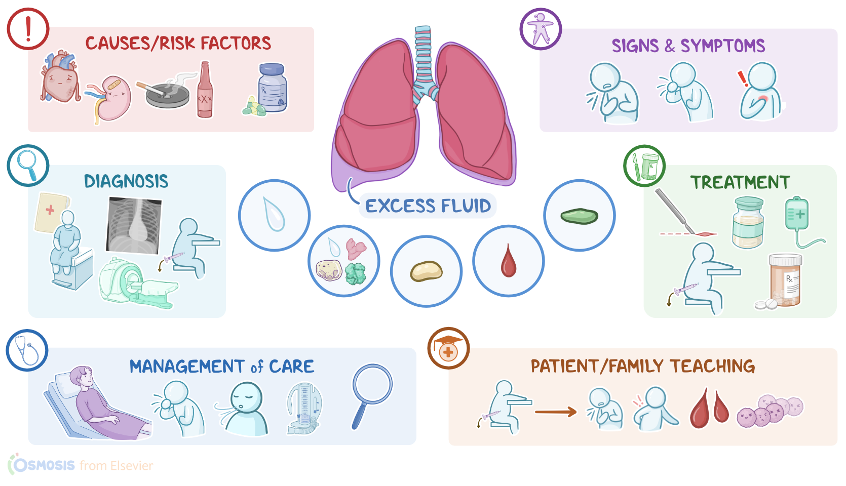
Pleural effusion is a condition in which excess fluid accumulates in the space between the visceral layer and parietal layer of the pleura, called the pleural space.

Normally, this space contains 20 to 25 milliliters of fluid that provides lubrication, allowing the two pleural layers to slide over each other during breathing. This pleural fluid forms as a filtrate from pleural blood vessels. At the same time, it is drained into the lymphatic vessels, and this allows for regular renewal of the fluid.

Now, pleural effusion is typically caused by increased production or impaired drainage of the pleural fluid. Depending on the cause, the excess fluid in pleural effusion can be protein-poor, called transudate, or protein-rich, called exudate.

Transudate, also called hydrothorax when it involves the pleural space, forms when too much fluid starts to move from the pulmonary capillaries into the pleural space, either because of increased hydrostatic pressure or decreased oncotic pressure within the pulmonary capillaries. So increased hydrostatic pressure occurs usually in the context of heart failure, where the heart can’t pump blood effectively, so it backs up into the pulmonary vessels, leading to pulmonary hypertension; ultimately, the high pressure forces fluid out of the pulmonary capillaries and into the pleural space. On the other hand, decreased oncotic pressure can be caused by cirrhosis, which leads to decreased hepatic production of plasma proteins like albumin; or nephrotic syndrome, where renal filtration of blood is impaired, so the proteins are lost in urine.

On the other hand, exudate forms when there’s increased permeability of the pulmonary capillaries, which allows fluid, immune cells, and large proteins, along with lactate dehydrogenase or LDH, to leak out of the capillaries and into the pleural space. This can be caused by trauma, malignancy, such as lung cancer, inflammatory conditions like pancreatitis, and systemic lupus erythematosus, or an infection like pneumonia.

Additionally, in some cases, the fluid could be purulent, meaning it contains pus, which is called an empyema or pyothorax, and is usually caused by bacterial infection like pneumonia; or the fluid could be sanguineous, meaning it contains blood, called a hemothorax, and it’s usually caused by blunt chest trauma, as well as malignancy, or pulmonary embolism; or it could contain lymph, which is called a chylothorax, and it can be caused by lymphatic system injury from trauma or medical procedures. So, risk factors for pleural effusion include underlying heart, lung, kidney, or liver conditions, as well as exposure to tobacco smoke, alcohol, and the use of certain medications, such as immunosuppressants.

Now, the excess fluid building up in the pleural space puts pressure on the lungs and its surrounding structures, like the trachea for example, which may cause impaired ventilation or oxygenation. In addition, impaired drainage of pleural fluid causes this fluid to stagnate, which allows bacteria to grow and cause infections.

Now, a small pleural effusion typically causes no clinical manifestations. With larger pleural effusions, though, clients may present with dyspnea, dry cough, and sharp pain that comes with inspiration. During auscultation, there are reduced or absent breath sounds on the affected side. On percussion, there is dullness due to the presence of fluid within the pleural space.

The diagnosis of pleural effusion starts with the client’s history and physical assessment, followed by a chest X-ray or CT scan to visualize the effusion. A large effusion might also show a collapsed lung.

Diagnostic thoracentesis is frequently also done, which is a procedure that involves the collection of fluid from the pleural space to determine its composition and find out its cause. The main difference is that exudative effusions are rich in protein or LDH, while transudative effusions aren’t. So to definitively determine if the fluid is exudative or transudative, the Light’s criteria can be used.

Pleural fluid is considered exudative if any one of following are met: a pleural fluid total protein to serum total protein ratio more than 0.5, a pleural fluid lactate dehydrogenase, or LDH, to serum LDH ratio more than 0.6, or a pleural fluid LDH more than two-thirds of the upper limit of normal serum LDH. It’s very important to not miss an exudative effusion, so the criteria are designed to be overly sensitive. Now if none of the Light’s criteria apply, it’s a transudative effusion. In that case, it’s okay to treat the underlying cause, and no further tests are needed.