Chest tube care: Nursing
A chest tube can be used to remove air, fluids, or blood from the pleural space; to prevent air or fluid from reentering the pleural space; and to reestablish intrapleural and intrapulmonary pressures after surgery or trauma, in order to re-expand the lungs. A chest tube can also be inserted into the mediastinum after open heart surgery to drain fluid from the pericardial sac. Now, let’s quickly review the physiology of the pleura, which is a two-layered serous membrane that covers the lungs. The first layer of the pleura adheres to the surface of the lungs, called visceral pleura, whereas the second layer lines the chest wall, and it’s called the parietal pleura. Between these two layers there’s the pleural space, which is typically filled with a small amount of serous fluid that prevents friction between the two layers while breathing.
Moreover, let’s look at the physiology of breathing, which consists of two stages: inspiration and expiration. Inspiration is an active process, meaning that it requires muscles such as the diaphragm and the intercostal muscles to contract, which increases the intrathoracic volume and lowers the intrapulmonary pressure below the atmospheric pressure. This pressure gradient makes air flow from a high pressure area, meaning the atmospheric air, to a lower pressure area; so air is pulled inside the lungs. On the other hand, expiration is a passive process, meaning that it doesn’t require muscle contraction. That’s because the lungs have a special feature called elastic recoil, meaning they have a tendency to return to their normal size after being expanded. This allows the lungs to deflate after inspiration, decreasing the intrathoracic volume and increasing the intrathoracic pressure.
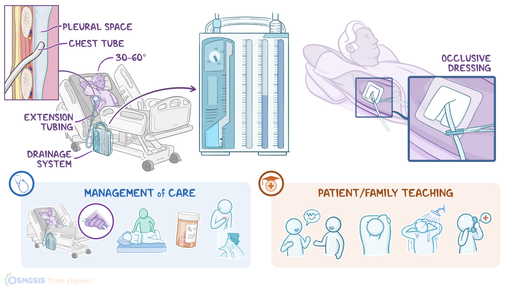
This process passively pushes the air from the area of higher pressure, which is the lungs, to the area of lower pressure, which is the atmosphere.Now, one end of the chest tube is typically inserted through the rib cage into the pleural space, while the other end is connected to extension tubing that drains into a drainage system. The most commonly used drainage system is the three-compartment drainage system. This system consists of three interconnected chambers: First, the collection chamber connects directly to the chest tube, and it collects air and fluid from the pleural cavity. Fluid stays in this chamber, while air flows to the next chamber, called the water seal chamber. This chamber acts as a one-way valve that allows air to flow into it during exhalation, and prevents air from moving back into the pleural space during inhalation. It is kept filled with 2 cm of sterile water that rises and falls during inhalation and exhalation, a process called tidaling.
| CHEST TUBE CARE | ||
| KEY POINTS | NOTES | |
| DEFINITION |
| |
| PHYSIOLOGY |
| |
| CHEST TUBES AND DRAINAGE SYSTEMS |
| |
| PROCEDURE |
| |
| MANAGEMENT OF CARE |
| |
| PATIENT AND FAMILY TEACHING |
| |
No notes for this video yet
Try adding a note below