Aortic Regurgitation · What Is It, Causes, Diagnosis, Treatment, and More

Published: Aug 01, 2025
Author: Michail Mavrogiannis, MD
Editor: Alyssa Haag, MD
Editor: Ian Mannarino, MD, MBA
Editor: Anna Hernández, MD
Illustrator: Jillian Dunbar
Copyeditor: Sadia Zaman, MBBS, Bsc
7-day free trial

Go deeper with Osmosis

Osmosis is a learning platform with videos, questions, and AI tools to help you master topics like this.

4.8 · 12,000+ reviews
Watch quick, visual videos
Practice with Qbank-style questions
Use AI to explain, quiz, and review
Study anytime with the mobile app
Start free trial

No credit card · Cancel anytime

What is aortic regurgitation?

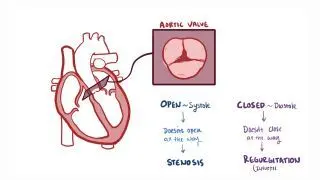
Aortic regurgitation, or aortic insufficiency, is a condition in which there is inadequate closure of the aortic valve during cardiac diastole, leading to the leakage of blood from the aorta into the left ventricle. As a result, there is reverse blood flow through the aortic valve.  

The aortic valve is typically made up of three leaflets and opens widely during systole to allow the ejection of blood, which is pumped from the left ventricle through the aorta to the rest of the body. During diastole, the aortic valve closes to allow the heart to fill with blood and prepare for the subsequent systole. If the valve doesn’t close completely, blood leaks back into the left ventricle, and aortic regurgitation occurs.  

Learn deeper with Osmosis

Master this topic faster with videos, questions, and AI.

Used by 8M+ healthcare learners.

Start free trial

No credit card · Cancel anytime

How common is aortic regurgitation?

Aortic regurgitation is a relatively common condition, affecting around 5-10% of the population. It occurs more commonly in individuals who are assigned male at birth. The prevalence of aortic regurgitation increases with age, and it occurs most often in individuals older than 50 years of age. 

What causes aortic regurgitation?

Aortic regurgitation is caused by issues with the supporting structures of the aortic root, which is the first part of the aorta leaving from the heart, or inadequate closure of the aortic valve leaflets during diastole. This can be either due to an impairment of the leaflets, or an increased afterload, which is the resistance that the heart needs to overcome to open the aortic valve.  

The most common cause of aortic regurgitation is aortic root dilation, in which the aortic root becomes enlarged. It mainly occurs as a result of aging. Other causes of aortic root dilation include various conditions, such as Behcet disease, reactive arthritis, Marfan syndrome, osteogenesis imperfecta, syphilitic aortitis, and aortic dissection 

A common cause of leaflet impairment includes aortic valve calcification. Aortic valve calcification can either result from normal aging or can be caused by congenital conditions, such as a bicuspid aortic valve, which can accelerate calcification. Other causes of leaflet impairment include infectious diseases, such as bacterial endocarditis (i.e., infective endocarditis) and rheumatic fever; inflammatory disorders such as systemic lupus erythematosus (SLE) and rheumatoid arthritis; and traumatic injury. High blood pressure, supravalvular aortic stenosis, and coarctation of the aorta (i.e., narrowing in the aorta) can also lead to aortic regurgitation by increasing the afterload. 

What are the signs and symptoms of aortic regurgitation?

In the early stages of the condition, individuals with aortic regurgitation are typically asymptomatic. As aortic regurgitation develops gradually, symptoms may take years or even decades to manifest. When they do occur, symptoms include exertional dyspnea (i.e., shortness of breath upon exercise), orthopnea, (i.e., shortness of breath upon lying), paroxysmal nocturnal dyspnea (i.e., shortness of breath at night when the heart rate slows), angina (i.e., chest pain), peripheral edema (i.e., swollen ankles and feet), fainting, heart murmur, and palpitations. Sometimes, aortic regurgitation can occur acutely, typically as a result of an infection of the aortic valve, resulting in sudden shortness of breath due to pulmonary edemawhere excess fluid accumulates in the lungs.  

Usually, the murmur aortic regurgitation produces is a characteristic whooshing sound that can be heard over the individual’s chest with the help of a stethoscope. The murmur can be best heard with the individual sitting up, leaning forward, and holding their breath in full expiration. These maneuvers bring the aortic valve closer to the chest wall and reduce lung sounds. 

Aortic regurgitation is also associated with classic physical examination findings that result from the consequences of the regurgitant blood flow, specifically a wide pulse pressure, which is the difference between systolic and diastolic pressure. As blood regurgitates back into the left ventricle during diastole, the diastolic pressure in the aorta decreases, leading to an increase in pulse pressure. Additionally, systolic blood pressure is often increased to compensate for the reduction in diastolic pressure, in an attempt to drive the forward flow of blood, which further widens the pulse pressure. Some of these classic clinical signs include the Becker sign, which is the visible pulsation of retinal blood vessels via an ophthalmoscope; Corrigan’s pulse, which is a rapid, forceful upstroke of the pulse followed by a sudden collapse; and the Musset sign, which is when the head bobs in sync with each heartbeat. 

What are the differential diagnoses for aortic regurgitation?

Differential diagnoses involve considering various possible conditions that could be causing symptoms and then ruling out each one through use of history, clinical evaluation, diagnostic tests, and critical thinking. This process helps to narrow down the list of potential diagnoses to determine the most likely cause of the symptoms.

Differential diagnoses can be broken down into four categories: most likely, less likely, least likely, and can’t miss. Most likely diagnoses are conditions most probable based on symptoms and clinical presentation. Less likely diagnoses are not as probable but should still be considered. On the other hand, least likely diagnoses can be considered if other, more probable conditions are excluded. Finally, can’t miss diagnoses are less common but critical to promptly identify and treat as they can lead to severe consequences.

Differential diagnoses for aortic regurgitation include:
Most likely:
- Bicuspid aortic valve: A congenital anomaly that predisposes to valve degeneration and regurgitation.
- Rheumatic heart disease: Can cause chronic valvular damage, especially in endemic areas.
- Infective endocarditis: Damages the valve leaflets, leading to acute or chronic regurgitation.

Less likely:
- Marfan syndrome or other connective tissue disorders: Cause aortic root dilation and valve incompetence.
- Ankylosing spondylitis: Can be associated with aortic root inflammation and regurgitation.

Least likely:
- Systemic lupus erythematosus (SLE): May involve the heart valves.
- Radiation-induced valvular disease: Seen in patients with prior mediastinal radiation therapy.

Can’t miss:
- Acute aortic dissection: Can cause sudden-onset severe aortic regurgitation and hemodynamic collapse.
- Infective endocarditis with valve destruction: Can lead to acute regurgitation and heart failure.
- Traumatic aortic valve rupture: Requires immediate recognition and intervention.
- Prosthetic valve dysfunction: In patients with prior valve replacement, malfunction or dehiscence can cause acute regurgitation.
How is aortic regurgitation diagnosed?

Aortic regurgitation is typically diagnosed after a thorough review of signs and symptoms and can be confirmed with an echocardiography, or an ultrasound of the heart. Findings from the echocardiography may include dilation of the left ventricle, which is commonly seen in chronic aortic regurgitation, as well as decreased ejection fraction (EF), which is the percentage of blood leaving the left ventricle with each heartbeatA chest x-ray may also help establish the diagnosis and may show an enlarged heart due to the enlargement of the left ventricle or signs of pulmonary edema. An EKG, or electrocardiogram, may indicate hypertrophy, or increases in the size of the heart muscle cells, of the left ventricle. Other imaging modalities, such as cardiac magnetic resonance (CMR), may be useful to measure the aortic root and assess the anatomy of the ascending aorta. Pulmonary artery catheterization, which is more invasive, may also help establish the severity of the condition if echocardiography findings are inconclusive.  

How is aortic regurgitation treated?

Management of individuals with aortic regurgitation depends on the signs and symptoms of the disease, the degree of the left ventricular dysfunction, and whether aortic regurgitation is  acute or chronic. Individuals with mild disease usually don’t need treatment and may be monitored annually to assess the progression of aortic regurgitation. Asymptomatic individuals may be recommended surgical treatment if heart function is impaired or if there is severe left ventricular dilation. Individuals with mild to severe aortic regurgitation who experience significant symptoms or show an abnormal response to exercise testing, usually undergo surgery. Surgical treatment, which involves aortic valve replacement (AVR) is the definitive treatment of aortic regurgitation. Medical treatment, which aims at reducing the afterload and relieving symptoms, can also be used and often includes ACE inhibitors, such as ramipril, and calcium channel blockers, such as amlodipine  

For individuals with acute aortic regurgitation emergencies, surgical treatment with aortic valve replacement is recommended to prevent life-threatening complications. Medications may also be used to stabilize the individual until surgery is performed.  

What are the most important facts to know about aortic regurgitation?

Aortic regurgitation, or aortic insufficiency, is a relatively common and serious condition in which there is inadequate closure of the aortic valve during diastole, causing reverse blood flow through the aortic valve and leakage of blood from the aorta into the left ventricle. The most common cause of aortic regurgitation is aortic root dilation, which is most commonly the result of aging. Leaflet impairment, which can either result from normal aging or congenital conditions such as bicuspid aortic valve, can also cause aortic regurgitation. Other causes include hypertension, which can lead to aortic regurgitation by increasing the afterload. The key tool for diagnosing aortic regurgitation is echocardiographyManagement of individuals with aortic regurgitation depends on the signs and symptoms of the disease, the degree of left ventricular dysfunction, and the duration of the disease. Aortic valve replacement (AVR) is the definitive treatment of aortic regurgitation for individuals who require treatment.  

Key Takeaways

Definition 

A condition characterized by inadequate closure of the aortic valve during cardiac diastole, leading to reverse blood flow from the aorta into the ventricle. 

Risk Factors 

- 5-10% of the population 

- More common in:  

     - Assigned males at birth  

     - Individuals older than 50 years of age 

Causes 

- Aortic root dilation  

- Aortic valve calcification 

- Bacterial endocarditis 

- Rheumatic fever 

- Systemic lupus erythematosus 

- Rheumatoid arthritis  

- Traumatic injury  

- High blood pressure 

- Supravalvular aortic stenosis 

- Aorta coarctation 

Signs and Symptoms 

- Early stage → asymptomatic. Symptoms develop over years or decades. 

- Exertional dyspnea 

- Orthopnea 

- Paroxysmal nocturnal dyspnea 

- Angina 

- Peripheral edema 

- Fainting 

- Heart murmur 

- Palpitations 

- Pulmonary edema  

Differential Diagnosis 

- Most likely:  

     - Bicuspid aortic valve 

     - Rheumatic heart disease 

     - Infective endocarditis  

- Can’t miss:  

     - Acute aortic dissection 

     - Infective endocarditis  

     - Traumatic aortic valve rupture 

     - Prosthetic valve dysfunction 

Diagnosis 

- Physical examination findings: 

     - Auscultation: whooshing sound best heard while sitting up, leaning forward, and holding breath in full expiration 

     - Wide pulse pressure  

     - Becker sign  

     - Corrigan’s pulse  

     - Musset sign 

- Review of signs and symptoms 

     - Echocardiography  

     - Chest X-ray  

     - EKG 

     - Cardiac magnetic resonance  

     - Pulmonary artery catheterization  

Treatment 

- Mild disease → annual monitoring to assess progression  

- Asymptomatic → surgery if heart function impaired or severe left ventricular dilation  

- Mild-severe disease + symptoms / abnormal response to exercise testing → surgery  

- Acute aortic regurgitation emergenciessurgical treatment 

- Surgical treatment: aortic valve replacement (AVR)  

- Medical treatment: ACE inhibitors, calcium channel blockers  

Students say Osmosis is 100% worth it

Because Osmosis saves them time. Lowers stress. And actually helps them remember when it counts.

I used Osmosis to prepare for my first medical school licensing exam! Super helpful and interactive for people who may not do great with just pages of text info!

Cecilia Ruiz

Cecilia Ruiz

MD student

Sayan Misra

I have used Osmosis for about four years. Best thing I have ever used for my medical studies.

Sayan Misra

Sayan Misra

Med student

Osmosis videos are superior because they define simple concepts, tell a story with a clear progression, and provide context.

Jay Pate

Jay Pate

Dental student

References


Akinseye OA, Pathak A, Ibebuogu UN. Aortic valve regurgitation: A comprehensive review. Curr Probl Cardiol. 2017;43(8):315-334. doi:10.1016/j.cpcardiol.2017.10.004 


Correction to: 2020 ACC/AHA guideline for the management of patients with valvular heart disease: A report of the American college of cardiology/American heart association joint committee on clinical practice guidelines. Circulation. 2024;150(12):e267. doi:10.1161/CIR.0000000000001284 


Marigliano AN, Ortiz JT, Casas J, Evangelista A. Aortic regurgitation: From valvular to myocardial dysfunction. J Clin Med. 2024;13(10):2929. doi:10.3390/jcm13102929