
Arachnoid Cysts · What Are They, Causes, Symptoms, and More

Published: Mar 30, 2026
Author: Nikol Natalia Armata, MD
Editor: Alyssa Haag, MD
Editor: Emily Miao, MD, PharmD
Editor: Kelsey LaFayette, DNP, ARNP, FNP-C
Illustrator: Abbey Richard, MSc
7-day free trial

Go deeper with Osmosis

Osmosis is a learning platform with videos, questions, and AI tools to help you master topics like this.

4.8 · 12,000+ reviews
Watch quick, visual videos
Practice with Qbank-style questions
Use AI to explain, quiz, and review
Study anytime with the mobile app
Start free trial

No credit card · Cancel anytime

What is an arachnoid cyst?

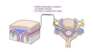
An arachnoid cyst refers to a type of spinal fluid-filled sac that forms between the brain or spinal cord and the arachnoid membrane, a protective layer that surrounds the central nervous system (i.e., brain and spinal cord). These cysts can be congenital, meaning they are present from birth, and are more commonly identified in those assigned males at birth; or can develop later in life due to trauma or infection. Most arachnoid cysts form near the temporal lobe of the brain in an area of the skull known as the middle cranial fossa. Arachnoid cysts involving the spinal cord are rarer. The location and size of the cyst determine the severity and quality of symptoms. 

Learn deeper with Osmosis

Master this topic faster with videos, questions, and AI.

Used by 8M+ healthcare learners.

Start free trial

No credit card · Cancel anytime

What causes an arachnoid cyst?

The exact cause of arachnoid cyst formation is not always clear, but they are generally believed to be congenital and rarely, acquired. 

One theory suggests that during fetal development, the arachnoid membrane may not properly fuse, leading to a small pouch or sac that fills with cerebrospinal fluid (CSF) and forms a cyst. Another possible cause is a developmental abnormality in the brain or spinal cord that leads to the formation of a cyst. 

Additionally, arachnoid cysts can also develop later in life due to situations that disrupt the normal flow or absorption of CSF leading to the accumulation of fluid within the arachnoid space and the formation of a cyst. Examples of situations that can lead to acquired cysts can include head trauma, after a lumbar puncture, or infection (e.g., meningitis). Nonetheless, the exact mechanisms underlying the formation of arachnoid cysts are still not fully understood and may vary depending on individual cases. 

What are the signs and symptoms of an arachnoid cyst?

Signs and symptoms of an arachnoid cyst can vary widely depending on factors such as the size and location of the cyst. In many cases, individuals may experience no symptoms at all, especially if the cyst is small and does not compress the surrounding structures. However, when symptoms do occur, they often include persistent or recurrent headaches, which may be localized to the area of the cyst, as well as weakness or paralysis, usually on one side of the body. Seizures can also occur in some individuals, ranging in severity. Increased pressure on the brain due to a large cyst may lead to nausea and vomiting. 

Additionally, cysts located in the cerebellum can affect coordination and stability, resulting in difficulties with balance and walking or maintaining posture. Visual disturbances may occur if the cyst is near the optic nerve or other visual pathways. In children, particularly if the cyst is large and causes increased pressure on the brain, there may be delays in cognitive development, meaning their ability to think and reason, or other neurological deficits. In some cases, an arachnoid cyst may obstruct the normal flow of CSF, leading to hydrocephalus, characterized by the accumulation of fluid within the brain's ventricles and increased pressure within the skull. 

How is an arachnoid cyst diagnosed?

Diagnosing arachnoid cysts typically involves a comprehensive approach that begins with a thorough medical history and physical examination, conducted to evaluate the individual’s neurological function and detect any signs of increased intracranial pressure (e.g., dilation of pupils, involuntary flexion or extension of the arms and legs, decreased level of consciousness). Imaging studies, particularly magnetic resonance imaging (MRI), are commonly used to visualize arachnoid cysts in detail. MRI provides high-resolution images of the brain and spinal cord, allowing healthcare providers to accurately identify the presence, size, location, and characteristics of the cyst. In emergencies or when MRI is not accessible, CT scans, or computed tomography, may be used as an alternative. A lumbar puncture, or spinal tap, may be performed to measure CSF pressure and assess for signs of increased intracranial pressure.  

How is an arachnoid cyst treated?

Arachnoid cyst treatment depends on various factors, including the size and location of the cyst, as well as the presence and severity of symptoms. In many cases, small, asymptomatic cysts may not require active treatment and can be monitored periodically with imaging tests to ensure they do not grow or cause problems over time. However, if the cyst is large, causes significant symptoms, or leads to complications such as hydrocephalus, treatment options may be considered. Minimally invasive interventions may be necessary to drain the cyst or remove it entirely, particularly if it is compressing vital structures or obstructing the flow of cerebrospinal fluid. Cystoperitoneal shunting is an invasive option often chosen for treatment, during which a catheter is implanted into the cyst within the brain. Connected through a one-way valve, it empties the excessive fluid into the peritoneal cavity in the abdomen. The purpose of the shunt is to drain excess CSF to another part of the body where it can be absorbed, thus relieving pressure on the brain.   

The available surgical approaches include endoscopic surgery (i.e., use of a thin, flexible endoscope to access a specific area of the brain through small incisions or natural openings, such as the nostrils) and craniotomy (i.e., removal of a section of the skull to temporarily access the brain). Once the cyst is accessed, depending on the individual case, various techniques may be performed such as cyst fenestration (i.e., creating a small opening in the cyst wall to allow fluid to drain) or cyst removal. The goal of treatment is to relieve symptoms, prevent complications, and improve overall quality of life.  

What are the most important facts to know about an arachnoid cyst?

Arachnoid cysts are fluid-filled formations between the brain or spinal cord and the arachnoid membrane. Most arachnoid cysts are present at birth, although some may develop later in life. Their location and size may vary, causing different symptoms, from being completely asymptomatic to causing intense headaches, weakness, visual disturbances, vomiting, instability, and seizures. The diagnosis of such cysts is based on any indicative signs and symptoms as well as MRI or CT imaging. Treating arachnoid cysts is not always necessary, but if the cyst causes any symptoms or complications, surgical interventions are chosen to open the cyst and drain it. 

Key Takeaways

Definition 

An arachnoid cyst refers to a type of spinal fluid-filled sac that forms between the brain or spinal cord and the arachnoid membrane. 

Location 

-Most common location: temporal lobe  

-Rarely: spinal cord  

-Location and size → severity and quality  

Causes 

-Congenital  

-Improper fusion of the arachnoid membrane 

-Developmental abnormalities  

-(Rarely) Acquired (disruption of normal CSF flow/absorption)  

-Trauma  

-Infection (e.g., meningitis 

Signs and symptoms 

-Small cysts: often asymptomatic  

-Persistent or recurrent headaches  

-Unilateral weakness or paralysis  

-Seizures  

-Nausea, vomiting  

-Difficulties with balance, walking, posture maintenance (cerebellar involvement)  

-Visual disturbances (visual pathway involvement)  

-Children: cognitive delays / neurological deficits 

-Hydrocephalus   

Diagnosis 

-Medical history 

-Physical examination  

-Imaging (MRI, CT scan)  

-Lumbar puncture  

Treatment 

-Small, asymptomatic cysts → periodic monitoring with imaging 

-Large, symptomatic cysts:  

-Drainage (minimally invasive)  

-Cystoperitoneal shunting  

-Surgery: access via endoscopic surgery or craniotomy 

-Cyst fenestration  

-Cyst removal 

Students say Osmosis is 100% worth it

Because Osmosis saves them time. Lowers stress. And actually helps them remember when it counts.

I used Osmosis to prepare for my first medical school licensing exam! Super helpful and interactive for people who may not do great with just pages of text info!

Cecilia Ruiz

Cecilia Ruiz

MD student

Sayan Misra

I have used Osmosis for about four years. Best thing I have ever used for my medical studies.

Sayan Misra

Sayan Misra

Med student

Osmosis videos are superior because they define simple concepts, tell a story with a clear progression, and provide context.

Jay Pate

Jay Pate

Dental student

References


Ahmed AK, Cohen AR. Intracranial arachnoid cysts. Childs Nerv Syst. 2023;39(10):2771-2778. doi:10.1007/s00381-023-06066-0 


Arachnoid Cyst Fenestration. Elsevier eBooks. Published online January 1, 2020:289-292. doi:https://doi.org/10.1016/b978-0-323-52381-3.00051-4


Fernando SM, Tran A, Cheng W, et al. Diagnosis of elevated intracranial pressure in critically ill adults: systematic review and meta-analysis. BMJ. 2019;366:l4225. Published 2019 Jul 24. doi:10.1136/bmj.l4225 


Merola J, Manivannan S, Ooi S, et al. The efficacy of cystoperitoneal shunting for the surgical management of intracranial arachnoid cysts in the elderly: A systematic review of the literature. Surg Neurol Int. 2021;12:624. Published 2021 Dec 20. doi:10.25259/SNI_463_2021 


Mustansir F, Bashir S, Darbar A. Management of arachnoid cysts: A comprehensive review. Cureus. 2018;10(4):e2458. Published 2018 Apr 10. doi:10.7759/cureus.2458  


Westermaier T, Schweitzer T, Ernestus RI. Arachnoid cysts. Adv Exp Med Biol. 2012;724:37-50. doi:10.1007/978-1-4614-0653-2_3