Epidural and subdural hematoma: Nursing

Last updated: June 17, 2022

Epidural and subdural hematoma: Nursing

Medical Surgical

Medical Surgical

Arrhythmias - Asystole: Nursing
Arrhythmias - Atrial fibrillation (Afib): Nursing
Arrhythmias - Atrial flutter (Aflutter): Nursing
Arrhythmias - Heart blocks: Nursing
Arrhythmias - Premature atrial contractions (PACs): Nursing
Arrhythmias - Premature ventricular contractions (PVCs): Nursing
Arrhythmias - Sinus tachycardia and sinus bradycardia: Nursing
Arrhythmias - Supraventricular tachycardia (SVT): Nursing
Arrhythmias - Ventricular fibrillation (Vfib): Nursing
Arrhythmias - Ventricular tachycardia (Vtach): Nursing
Arterial embolism: Nursing
Electrocardiogram (ECG) - Normal sinus rhythm (NSR): Nursing
Cardiomyopathy: Nursing
Congenital heart defects - Acyanotic: Nursing
Congenital heart defects - Cyanotic: Nursing
Endocarditis: Nursing
Heart defects that decrease pulmonary blood flow - Nursing considerations & client education: Nursing
Kawasaki disease: Nursing
Myocarditis: Nursing
Pericarditis: Nursing
Shock - Anaphylactic: Nursing
Shock - Cardiogenic: Nursing
Shock - Hypovolemic: Nursing
Shock - Neurogenic: Nursing
Shock - Obstructive: Nursing
Shock - Septic: Nursing
Valvular heart disease: Nursing
Geriatric considerations - Cardiac: Nursing
Aortic aneurysm: Nursing process (ADPIE)
Coronary artery disease (CAD) and angina pectoris: Nursing process (ADPIE)
Hypertension: Nursing process (ADPIE)
Left-sided heart failure: Nursing process (ADPIE)
Myocardial infarction (MI): Nursing process (ADPIE)
Pericardial effusion and cardiac tamponade: Nursing process (ADPIE)
Rheumatic heart disease: Nursing process (ADPIE)
Alpha-1 adrenergic blockers: Nursing pharmacology
Alpha-2 adrenergic agonists: Nursing pharmacology
Angiotensin II receptor blockers (ARBs): Nursing pharmacology
Angiotensin-converting enzyme (ACE) inhibitors: Nursing pharmacology
Antiarrhythmics: Nursing pharmacology
Antihyperlipidemics - Fibrates: Nursing pharmacology
Antihyperlipidemics - Miscellaneous: Nursing pharmacology
Antihyperlipidemics - Statins: Nursing pharmacology
Beta-adrenergic blockers: Nursing pharmacology
Calcium-channel blockers: Nursing pharmacology
Cardiac glycosides: Nursing pharmacology
Direct-acting vasodilators: Nursing pharmacology
Nitrates: Nursing pharmacology
Sympathomimetic medications: Nursing pharmacology
Adrenal insufficiency (Addison disease): Nursing
Cushing syndrome and Cushing disease: Nursing
Hyperparathyroidism: Nursing
Hypoparathyroidism: Nursing
Hypopituitarism: Nursing
Infant of a diabetic mother (IDM): Nursing
Phenylketonuria (PKU): Nursing
Diabetes insipidus: Nursing process (ADPIE)
Diabetes mellitus (DM): Nursing process (ADPIE)
Diabetic ketoacidosis (DKA): Nursing process (ADPIE)
Hyperosmolar hyperglycemic state (HHS): Nursing process (ADPIE)
Hyperthyroidism: Nursing process (ADPIE)
Hypothyroidism: Nursing process (ADPIE)
Syndrome of inappropriate antidiuretic hormone (SIADH): Nursing process (ADPIE)
Glucocorticoids and mineralocorticoids: Nursing pharmacology
Insulin: Nursing pharmacology
Medications affecting the parathyroid glands: Nursing pharmacology
Medications for antidiuretic hormone (ADH) disorders: Nursing pharmacology
Medications for thyroid disorders: Nursing pharmacology
Medications for growth hormone disorders: Nursing pharmacology
Oral antidiabetic medications - Alpha-glucosidase inhibitors: Nursing pharmacology
Oral antidiabetic medications - Biguanides and thiazolidinediones: Nursing pharmacology
Oral antidiabetic medications - DPP-4 inhibitors: Nursing pharmacology
Oral antidiabetic medications - Sodium-glucose co-transporter-2 (SGLT-2) inhibitors: Nursing pharmacology
Oral antidiabetic medications - Sulfonylureas and meglitinides: Nursing pharmacology
Amblyopia: Nursing
Cataracts: Nursing
Detached retina: Nursing
Hearing impairment and otosclerosis: Nursing
Legal blindness: Nursing
Macular degeneration: Nursing
Meniere disease: Nursing
Pharyngitis: Nursing
Strabismus: Nursing
Geriatric considerations - Sensory: Nursing
Otitis media: Nursing
Epistaxis: Nursing process (ADPIE)
Eye injury: Nursing process (ADPIE)
Glaucoma: Nursing process (ADPIE)
Tonsillitis: Nursing process (ADPIE)
Antiglaucoma medications: Nursing pharmacology
Eye anesthetics: Nursing pharmacology
Mydriatics and cycloplegics: Nursing pharmacology
Ophthalmic anti-inflammatories and anti-infectives: Nursing pharmacology
Biliary atresia: Nursing
Cholecystitis: Nursing
Cholelithiasis: Nursing
Diarrhea: Nursing
Diverticular disease: Nursing
Hepatitis: Nursing
Inflammatory bowel disease - Crohn disease and ulcerative colitis: Nursing
Intestinal obstruction: Nursing
Irritable bowel syndrome (IBS): Nursing
Jaundice: Nursing
Esophageal atresia and tracheoesophageal fistula: Nursing
Geriatric considerations - Gastrointestinal: Nursing
Hirschsprung disease: Nursing
Hyperemesis gravidarum: Nursing
Necrotizing enterocolitis: Nursing
Omphalocele and gastroschisis: Nursing
Appendicitis: Nursing process (ADPIE)
Celiac disease: Nursing process (ADPIE)
Cirrhosis: Nursing process (ADPIE)
Gastroesophageal reflux disease (GERD): Nursing process (ADPIE)
Hiatal hernia: Nursing process (ADPIE)
Pancreatitis: Nursing process (ADPIE)
Peptic ulcer disease (PUD): Nursing process (ADPIE)
Antacids: Nursing pharmacology
Antidiarrheals: Nursing pharmacology
Antiemetics: Nursing pharmacology
Antispasmodics (GI): Nursing pharmacology
Gallstone-dissolving agents: Nursing pharmacology
Gastric mucosal protective agents: Nursing pharmacology
Histamine H2 antagonists: Nursing pharmacology
Laxatives: Nursing pharmacology
Medications for hepatic encephalopathy: Nursing pharmacology
Pancreatic enzyme replacements: Nursing pharmacology
Proton pump inhibitors (PPIs): Nursing pharmacology
Treatment for Helicobacter pylori: Nursing pharmacology
Weight loss medications: Nursing pharmacology
Chronic kidney disease (CKD): Nursing
Dialysis care: Nursing
Epididymitis: Nursing
Glomerulonephritis: Nursing
Nephrotic syndrome: Nursing
Polycystic kidney disease (PKD): Nursing
Pyelonephritis: Nursing
Renal and urinary calculi: Nursing
Urinary retention: Nursing
Bladder exstrophy: Nursing
Circumcision: Nursing
Cryptorchidism: Nursing
Enuresis: Nursing
Geriatric considerations - Urinary: Nursing
Hypospadias and epispadias: Nursing
Acute kidney injury (AKI): Nursing process (ADPIE)
Benign prostatic hyperplasia (BPH): Nursing process (ADPIE)
Urinary incontinence - Stress: Nursing process (ADPIE)
Urinary tract infections (UTIs): Nursing process (ADPIE)
Diuretics - Osmotic and carbonic anhydrase inhibitors: Nursing pharmacology
Diuretics - Thiazide, thiazide-like, loop, and potassium-sparing diuretics: Nursing pharmacology
Antispasmodics (GU): Nursing pharmacology
Cholinergic therapy (GU): Nursing pharmacology
Anemia - Aplastic: Nursing
Anemia - Iron-deficiency: Nursing
Anemia - Macrocytic: Nursing
Disseminated intravascular coagulation (DIC): Nursing
Neutropenia: Nursing
Polycythemia: Nursing
Thalassemia: Nursing
Thrombocytopenia: Nursing
Arterial blood gas (ABG) - Overview: Nursing
Arterial blood gas (ABG) - Metabolic acidosis: Nursing
Arterial blood gas (ABG) - Respiratory alkalosis: Nursing
Blood cultures: Nursing
Cardiac biomarkers - Creatine kinase (CK): Nursing
Cardiac biomarkers - Troponin: Nursing
Coagulation studies - Partial thromboplastin time (PTT): Nursing
Complete blood count (CBC) - Hemoglobin and hematocrit: Nursing
Complete blood count (CBC) - Platelets: Nursing
Complete blood count (CBC) - Red blood cells (RBC): Nursing
Complete blood count (CBC) - White blood cells (WBC) and differential: Nursing
Complete metabolic panel (CMP) - Blood urea nitrogen (BUN) and creatinine (Cr): Nursing
Complete metabolic panel (CMP) - Chloride: Nursing
Complete metabolic panel (CMP) - Estimated glomerular filtration rate (eGFR): Nursing
Complete metabolic panel (CMP) - Glucose: Nursing
Complete metabolic panel (CMP) - Liver function tests (LFT): Nursing
Complete metabolic panel (CMP) - Potassium: Nursing
Complete metabolic panel (CMP) - Sodium: Nursing
Hemolytic disease of the fetus and newborn: Nursing
Hemolytic uremic syndrome: Nursing
Hemophilia: Nursing process (ADPIE)
Leukemia: Nursing process (ADPIE)
Sickle cell disease: Nursing process (ADPIE)
Anticoagulants - Direct thrombin and factor Xa inhibitors: Nursing pharmacology
Anticoagulants - Heparin: Nursing pharmacology
Anticoagulants - Warfarin: Nursing pharmacology
Antihyperlipidemics - Bile acid sequestrants and cholesterol absorption inhibitors: Nursing pharmacology
Antiplatelet agents: Nursing pharmacology
Blood products: Nursing pharmacology
Hematopoietic growth factors: Nursing pharmacology
Hemostatics: Nursing pharmacology
Iron preparations: Nursing pharmacology
Thrombolytics: Nursing pharmacology
Immune response - Adaptive: Nursing
Autoimmunity: Nursing
Human immunodeficiency virus (HIV) and acquired immunodeficiency syndrome (AIDS): Nursing
Hypersensitivity reactions - Type I: Nursing
Hypersensitivity reactions - Type II: Nursing
Hypersensitivity reactions - Type III: Nursing
Hypersensitivity reactions - Type IV: Nursing
Inflammatory process: Nursing
Immune response - Innate: Nursing
Immunodeficiency disorders - Primary: Nursing
Scleroderma: Nursing
Immunodeficiency disorders - Secondary: Nursing
Sjögren syndrome: Nursing
Systemic lupus erythematosus (SLE): Nursing
Toxic shock syndrome (TSS): Nursing
Erythema infectiosum (Fifth disease): Nursing
Fever: Nursing
Infectious mononucleosis: Nursing
Mumps (Parotitis): Nursing
Neonatal sepsis: Nursing
Pertussis: Nursing
Poliomyelitis: Nursing
Postpartum infections: Nursing
Roseola (Exanthem subitum): Nursing
Rubella (German measles): Nursing
Rubeola (Measles): Nursing
Smallpox: Nursing
Zika virus: Nursing
Anaphylaxis: Nursing process (ADPIE)
Lyme disease: Nursing process (ADPIE)
Rheumatoid arthritis (RA): Nursing process (ADPIE)
Antirejection immunosuppressants: Nursing pharmacology
Biologic agents: Nursing pharmacology
Vaccines: Nursing pharmacology
Immunoglobulins: Nursing pharmacology
Immunosuppressants for autoimmune diseases: Nursing pharmacology
Immunomodulators: Nursing pharmacology
Disease-modifying therapy for multiple sclerosis: Nursing pharmacology
Acne: Nursing
Animal and snake bites: Nursing
Burn injury: Nursing
Cutaneous fungal infections: Nursing
Erysipelas and cellulitis: Nursing
Herpes simplex virus (HSV): Nursing
Herpes zoster: Nursing
Impetigo: Nursing
Insect stings and bites: Nursing
Pediculosis and scabies: Nursing
Psoriasis: Nursing
Rocky Mountain spotted fever (RMSF): Nursing
Skin cancer - Basal cell carcinoma, squamous cell carcinoma, and melanoma: Nursing
Urticaria: Nursing
Geriatric considerations - Integumentary: Nursing
Atopic dermatitis: Nursing process (ADPIE)
Frostbite: Nursing process (ADPIE)
Methicillin-resistant Staphylococcus aureus (MRSA): Nursing process (ADPIE)
Pressure injury: Nursing process (ADPIE)
Debridement agents: Nursing pharmacology
Keratolytics: Nursing pharmacology
Antibiotics - Topical: Nursing pharmacology
Antifungals - Topical: Nursing pharmacology
Corticosteroids - Topical: Nursing pharmacology
Medications for acne vulgaris: Nursing pharmacology
Amputation: Nursing
Carpal tunnel syndrome: Nursing
Herniated intervertebral disc: Nursing
Hip fractures: Nursing
Muscular dystrophies - Duchenne and Becker: Nursing
Myasthenia gravis: Nursing
Osteoarthritis: Nursing
Osteomyelitis: Nursing
Osteoporosis: Nursing
Paget disease of bone: Nursing
Craniosynostosis: Nursing
Developmental dysplasia of the hip: Nursing
Juvenile idiopathic arthritis: Nursing
Scoliosis: Nursing
Acute compartment syndrome: Nursing process (ADPIE)
Fractures: Nursing process (ADPIE)
Gout: Nursing process (ADPIE)
Musculoskeletal injuries: Nursing process (ADPIE)
Acetylcholinesterase inhibitors for myasthenia gravis: Nursing pharmacology
Analgesics: Nursing pharmacology
Skeletal muscle relaxants: Nursing pharmacology
Altered level of consciousness (LOC): Nursing
Amyotrophic lateral sclerosis (ALS): Nursing
Bell palsy: Nursing
Cerebral palsy: Nursing
Encephalitis: Nursing
Guillain-Barré syndrome: Nursing
Head injury: Nursing
Hemorrhagic stroke - Intracranial hemorrhage (ICH) and subarachnoid hemorrhage (SAH): Nursing
Epidural and subdural hematoma: Nursing
Huntington disease: Nursing
Increased intracranial pressure (ICP): Nursing
Intracranial aneurysm: Nursing
Migraines: Nursing
Multiple sclerosis (MS): Nursing
Physiology of pain: Nursing
Spinal cord injury (SCI): Nursing
Delirium: Nursing
Dementia: Nursing
Brachial plexus injury: Nursing
Neurological assessment - Neonate: Nursing
Neural tube defects: Nursing
Meningitis: Nursing process (ADPIE)
Parkinson disease: Nursing process (ADPIE)
Seizure disorder: Nursing process (ADPIE)
Stroke: Nursing process (ADPIE)
Trigeminal neuralgia: Nursing process (ADPIE)
Antiepileptics: Nursing pharmacology
Medications for Alzheimer disease: Nursing pharmacology
Medications for migraines: Nursing pharmacology
Biology of cancer: Nursing
Bladder tumors: Nursing
Bone tumors: Nursing
Brain tumors: Nursing
Cervical cancer: Nursing
Colorectal cancer: Nursing
Esophageal cancer: Nursing
Gastric cancer: Nursing
Laryngeal cancer: Nursing
Liver cancer: Nursing
Lung cancer: Nursing
Lymphoma - Hodgkin and non-Hodgkin: Nursing
Multiple myeloma: Nursing
Neuroblastoma: Nursing
Ovarian cancer: Nursing
Pancreatic cancer: Nursing
Prostate cancer: Nursing
Renal cancer: Nursing
Retinoblastoma: Nursing
Testicular cancer: Nursing
Breast cancer: Nursing process (ADPIE)
Tumor lysis syndrome (TLS): Nursing Process (ADPIE)
Alkylating agents: Nursing pharmacology
Angiogenesis inhibitors: Nursing pharmacology
Antimetabolites: Nursing pharmacology
Antitumor antibiotics: Nursing pharmacology
Hormones and hormone modulators for cancer: Nursing pharmacology
Other antineoplastics: Nursing pharmacology
Plant extracts for chemotherapy: Nursing pharmacology
Platinum-based agents: Nursing pharmacology
Buerger disease: Nursing
Raynaud phenomenon: Nursing
Peripheral arterial disease (PAD): Nursing process (ADPIE)
Peripheral venous disease (PVD): Nursing process (ADPIE)
Contraception - Barrier methods: Nursing
Contraception - Hormonal methods: Nursing
Contraception - Natural methods: Nursing
Contraception - Permanent methods: Nursing
Endometriosis: Nursing
Genital warts: Nursing
Infertility: Nursing
Syphilis: Nursing
Gestational trophoblastic disease: Nursing
Precocious puberty: Nursing
Candidiasis: Nursing process (ADPIE)
Gonorrhea and chlamydia: Nursing process (ADPIE)
Pelvic inflammatory disease (PID): Nursing process (ADPIE)
Acute respiratory distress syndrome (ARDS): Nursing
Anthrax: Nursing
Aspergillosis: Nursing
Atelectasis: Nursing
Chest tube care: Nursing
COVID-19: Nursing
Cystic fibrosis: Nursing
Flail chest: Nursing
Influenza: Nursing
Pleural effusion: Nursing
Pleurisy: Nursing
Pneumothorax and hemothorax: Nursing
Pulmonary contusion: Nursing
Pulmonary edema: Nursing
Rib fracture: Nursing
Rupture of diaphragm: Nursing
Sarcoidosis: Nursing
Tuberculosis (TB): Nursing
Bronchopulmonary dysplasia (BPD): Nursing
Congenital diaphragmatic hernia: Nursing
Geriatric considerations - Respiratory: Nursing
Neonatal respiratory distress syndrome (NRDS): Nursing
Asthma: Nursing process (ADPIE)
Bacterial pneumonia: Nursing process (ADPIE)
Bronchiolitis and respiratory syncytial virus (RSV): Nursing process (ADPIE)
Carbon monoxide poisoning: Nursing process (ADPIE)
Chronic obstructive pulmonary disease (COPD): Nursing process (ADPIE)
Epiglottitis: Nursing process (ADPIE)
Foreign body aspiration and upper airway obstruction: Nursing process (ADPIE)
Laryngotracheobronchitis (LTB) and croup: Nursing process (ADPIE)
Smoke inhalation injury: Nursing process (ADPIE)
Venous thromboembolism (VTE): Nursing process (ADPIE)
Antihistamines: Nursing pharmacology
Bronchodilators: Nursing pharmacology
Corticosteroids - Inhaled: Nursing pharmacology
Mast cell stabilizers - Inhaled: Nursing pharmacology
Leukotriene modifiers: Nursing pharmacology
Medications to control airway secretions: Nursing pharmacology
Oxygen therapy: Nursing pharmacology
Respiratory stimulants: Nursing pharmacology
Preoperative care: Nursing
Postoperative care: Nursing
Palliative and hospice care: Nursing
Postmortem care and considerations: Nursing

Notes

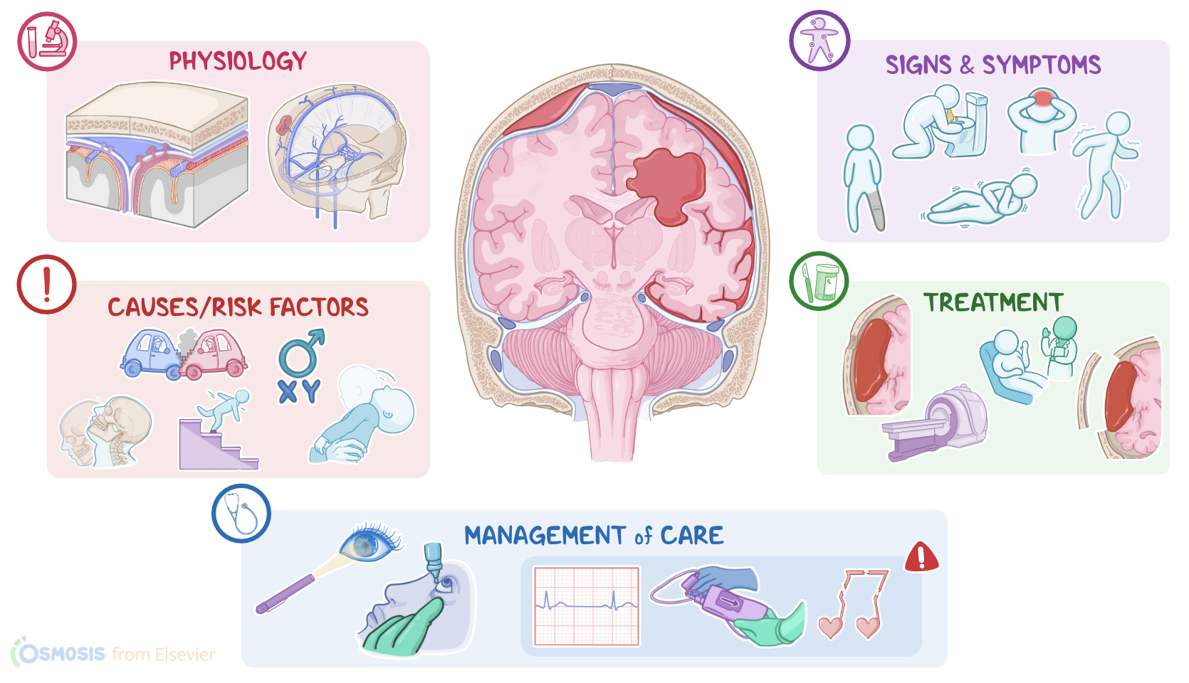
EPIDURAL AND SUBDURAL HEMATOMA

KEY POINTS
NOTES
DEFINITION
  • Intracranial hemorrhage: bleeding inside the skull
    • Epidural hematoma
      • Blood in epidural space
    • Subdural hematoma
      • Blood in subdural space
    • Subarachnoid hemorrhage
    • Intracerebral bleeding

PHYSIOLOGY
  • Meninges
    • Wrap around brain
    • Provide protection
    • Dura mater
      • Internal and external layer
        • Subdural space between internal layer and arachnoid mater
      • Meningeal arteries
    • Arachnoid mater
    • Subarachnoid space
      • Cerebrospinal fluid (CSF)
    • Pia mater

CAUSES AND RISK FACTORS
  • Causes - Epidural hematoma
    • Rupture of meningeal artery
      • Trauma
      • Tumors
      • Vascular malformations
      • Infection
      • Coagulation disorders
  • Risk factors - Epidural hematoma
    • Assigned male at birth
    • 20-30 years
  • Causes - Subdural hematoma
    • Rupture of veins in subdural space
      • Trauma
      • Shaken baby syndrome
  • Risk factors - Subdural hematoma
    • <1 year of age
    • Elderly
    • Chronic alcohol use
    • Neurodegenerative conditions

PATHOPHYSIOLOGY
  • Epidural hematoma
    • Meningeal artery tears
    • Blood pools between skull and external dura mater
    • ICP increases
  • Subdural hematoma
    • Vein tears
    • Blood pools in subdural space
    • ICP increases more slowly
  • Complications
    • Papilledema
    • Brain herniation

SIGNS AND SYMPTOMS
  • Epidural hematoma
    • Loss of consciousness
    • Can have period of lucidity followed by deterioration
    • Confusion
    • Coma
  • Subdural hematoma
    • Brief loss of consciousness 
    • Can have lucid period over longer period of time
  • Headaches
  • Seizures
  • Vomiting
  • Focal neurological symptoms
  • Signs of increased ICP
    • Cushing triad

DIAGNOSIS
  • History
  • Physical assessment
  • CT

TREATMENT
  • Surgery
  • CT
  • Monitoring
  • Drainage
  • Oxygen

MANAGEMENT OF CARE
  • Goals of care
    • Promote cerebral perfusion and oxygenation
    • Monitor for complications
  • Elevate head of bed
  • Maintain neutral head position
  • Create calm environment
  • Administer medications as prescribed
  • Monitor vital signs
  • Institute cardiac monitoring
  • Notify HCP
    • Cushing triad
    • Dysrhythmias
    • Decreased oxygen saturation
  • Routine postoperative care
  • Monitor for postoperative complications
  • Hyperoxygenate before suctioning
  • Check pupillary reaction
  • Maintain body temperature
  • Institute seizure prophylaxis
  • Maintain cerebral perfusion pressure
  • Perform neurovascular checks
    • Notify HCP
      • Sluggish pupils
      • Decreasing Glasgow Coma Scale score
  • Review laboratory results
    • Notify HCP
      • Hyperkalemia
      • Indications of diabetes insipidus
  • Watch surgical site
  • Keep drains dependent
    • Notify HCP
      • Drainge > 50mL in 8 hours

PATIENT AND FAMILY TEACHING
  • Explain condition, plan of care, and how to safely administer medications
  • Avoid medications that impact clotting
  • Emphasize rehab
  • Keep follow-up appointments
  • Lifestyle modifications
  • Safety precautions
  • Notify HCP
    • Drowsiness
    • Headache
    • Visual changes
    • Nausea
    • Vomiting
    • Behavioral changes

Transcript

Watch video only

Intracranial hemorrhage or ICH for short, refers to bleeding inside the skull. Types of intracranial hemorrhage include epidural hematoma, subdural hematoma, subarachnoid hemorrhage, and intracerebral bleeding. Now, epidural hematoma is the collection of blood in the epidural space, which is the space between the dura mater and inner surface of the skull. In contrast, subdural hematoma is the collection of blood in the subdural space, meaning between the dura mater and the arachnoid mater.

Let’s start by looking at the physiology of the meninges. This is a membrane that wraps around the brain and protects it from the outside environment. It’s made up of three meningeal layers. From outside to inside, these are the dura mater, arachnoid mater, and pia mater. Between the arachnoid and pia mater there’s the subarachnoid space, which houses the cerebrospinal fluid. The cerebrospinal fluid is a clear, watery liquid that cushions the brain from impact and bathes it in nutrients.

Now, the dura mater consists of an external and an internal layer. The external layer of the dura mater adheres tightly to the inner surface of the skull, so the epidural space is only a virtual space in the cranium, meaning the space isn’t appreciable unless pathology is present.

Between the external layer of the dura mater and the inner surface of the skull, there are meningeal arteries that supply meninges. The internal layer of the dura mater lies above the arachnoid mater, the two are separated by the subdural space. The subdural space plays a major role in venous blood drainage in the brain. The dura mater also forms dural venous sinuses which carry the venous blood from the brain. These sinuses are connected to the veins outside of the cranium by bridging veins.

Okay, let’s move onto epidural and subdural hematoma. The most common cause of epidural hematoma is rupture of the meningeal artery. This is often caused by blunt head trauma from falls or car crashes. The most common site for epidural hematoma is at the pterion which is the thinnest part of the skull, and it’s located right above the middle meningeal artery. Other causes of epidural hematoma may include hemorrhagic tumors, vascular malformations, infection or coagulation disorders. Risk factors for epidural hematoma include being assigned male at birth and age from 20 to 30 because the external layer of the dura mater hasn’t fully attached to the skull yet.

On the other hand, the main cause of a subdural hematoma is rupture of bridging veins located in the subdural space. Now, like with epidural hematoma, blunt head trauma is a major cause, but another important cause is acceleration-deceleration injury. This can occur when you step on the brakes while driving very fast, causing the head and brain to move forward and back violently, damaging the bridging veins. Another cause is shaken baby syndrome where a baby is violently shaken, making their head whip back and forth damaging the bridging veins.

Now, clients younger than 1 year of age and elderly clients are at higher risk because they often have relatively thin walled blood vessels that are susceptible to rupture. Old age, chronic alcohol use and neurodegenerative conditions like Alzheimer disease cause brain atrophy. When the brain shrinks, there’s more space around the bridging veins, leaving them exposed and unsupported.

Okay, moving on to pathology, with epidural hematoma once the meningeal artery is torn, blood will pool between the skull and the external layer of the dura mater. Now, what makes epidural hematomas so dangerous is that the dura mater is attached tightly to the sutures of the skull, so the blood can’t cross these sutures and becomes trapped. This can quickly increase the intracranial pressure or ICP and a large epidural hematoma on one side of the skull can cause a midline shift and push the brain towards the opposite side of the skull.

In the case of subdural hematoma, once a bridging vein is torn, the blood pools in the subdural space creating a hematoma. Because the source of the bleeding is venous, subdural hematomas usually grow slower than epidural hematomas which are caused by arterial bleeding. Also, since the blood isn’t restricted by sutures, it can be distributed over a larger area, so pressure doesn’t build up as quickly, unless the hematoma is very large.

Now, with both epidural and subdural hematoma, as blood accumulates over time the hematoma grows in size and increases the ICP. If not corrected, increased ICP can lead to complications, such as papilledema, which is the swelling of the optic disc where the optic nerve leaves the retina. The most severe complication, though, is brain herniation. Brain herniation occurs when a part of the brain is pushed into another space of the skull or even out of it through the foramen magnum, and this can affect other CNS structures, like the brainstem.

Clinical manifestations of an epidural hematoma most commonly include a loss of consciousness right after the head trauma. In a slow epidural bleed, this might be followed by a lucid interval of temporary improvement right after the initial trauma, but then hours later, they will deteriorate as the epidural hematoma grows and progresses to confusion and coma.

In the case of a subdural hematoma, there can also be a brief loss of consciousness followed by a lucid interval. However, depending on the size and speed of the bleed, this can last from days to weeks before symptoms develop. A subdural hematoma is considered acute if symptoms develop within 2 days of a head trauma, subacute if they develop between 2 days and 2 weeks of a head trauma, and chronic if they develop 2 weeks or more after a head trauma. Although symptom onset is slower than epidural hematoma, it’s actually more deadly since the blood can spread across a larger area around the brain, so deterioration can be quick once symptoms develop.

In both epidural and subdural hematomas, there can also be headaches, bouts of seizures and vomiting, as well as focal neurological symptoms like muscle weakness or sensory problems depending on the location of the hematoma. Additionally, there can be signs of increased ICP such as vomiting, a fixed dilated pupil on the same side as the hematoma, as well as the Cushing triad, which includes bradycardia, irregular respiratory pattern, and widening of pulse pressure where systolic pressure increase more than diastolic pressure.

Diagnosis of increased ICP starts with the client’s history and physical assessment, followed by a CT scan to confirm the diagnosis. In the case of an epidural hematoma, this typically shows a convex, “lens-shaped” collection of blood that does not cross the suture lines of the skull. In contrast, a subdural hematoma typically appears as a concave, “crescent-shaped” density that crosses the suture lines.

Once the diagnosis is confirmed, the client should be immediately sent to OR for surgery. Most epidural hematomas are treated by craniotomy, which is when part of the skull bone is removed in order to remove accumulated blood clot below. In some settings, a life-saving alternative is to drill a hole in the skull, which is called Burr hole trephination. Finally, non-surgical management with serial neurological assessments and CT scans might be considered for clients with small epidural hematomas and in good clinical condition.