Arthrocentesis · Indications, How It’s Performed, and More

Published: Mar 30, 2026
Author: Lily Guo, MD
Editor: Alyssa Haag, MD
Editor: Ian Mannarino, MD, MBA
Editor: Kelsey LaFayette, DNP, ARNP, FNP-C
Illustrator: Abbey Richard, MSc
7-day free trial

Go deeper with Osmosis

Osmosis is a learning platform with videos, questions, and AI tools to help you master topics like this.

4.8 · 12,000+ reviews
Watch quick, visual videos
Practice with Qbank-style questions
Use AI to explain, quiz, and review
Study anytime with the mobile app
Start free trial

No credit card · Cancel anytime

What is arthrocentesis?

Arthrocentesis, also known as joint aspiration, refers to the medical procedure whereby a needle is used to aspirate synovial fluid from a joint. Examples include knee arthrocentesis where fluid is removed from the knee joint or other joint arthrocentesis (i.e., hip. elbow, shoulder).  

Learn deeper with Osmosis

Master this topic faster with videos, questions, and AI.

Used by 8M+ healthcare learners.

Start free trial

No credit card · Cancel anytime

What are indications for arthrocentesis?

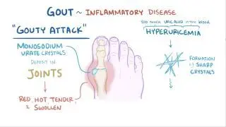
Arthrocentesis can be performed for both diagnostic purposes and for treatment, including pain relief. For example, arthrocentesis can help diagnosis joint effusion, or build-up of fluid, caused by rheumatoid arthritis, septic arthritis, hemorrhage resulting in hemarthrosis, or gout and pseudogoutRheumatoid arthritis is a chronic autoimmune disorder resulting in a swollen and painful joint, most commonly in the hands and wrists. Septic arthritis (i.e., a joint infection) can be categorized into gonococcal arthritis and nongonococcal arthritis. Nongonococcal arthritis is commonly caused by gram-positive staphylococci and streptococci, which is much more common than gonococcal arthritis. Septic arthritis commonly affects the knees, hips, and shoulders. Gout is a condition marked by the build-up of uric acid crystals in the body, commonly affecting the big toe. A large effusion in a small joint space can result in excess pressure and pain. Performing an arthrocentesis can help remove extra fluid and allow for rapid pain relief. After, the injection of medications (e.g., corticosteroids) can further help alleviate pain and inflammation.  

How is an arthrocentesis performed?

Most commonly, an arthrocentesis is performed by the clinician in the emergency department, hospital, or outpatient setting. Before performing the procedure, the clinician will palpate and define the joint anatomy by using bony landmarks to accurately identify the needle insertion site. Portable ultrasound can also be used to locate the effusion and determine its size and extent. Arthrocentesis is then performed under sterile conditions. First, the skin around the affected joint is cleaned with solutions such as chlorhexidine or betadine. The clinician then drapes the field with sterile drapes and puts on sterile gloves. Local anesthetic (e.g., lidocaine) is injected into the site to help with pain control. A syringe is attached to a needle, typically 18 to 22 gauge, and inserted into the joint space. The fluid is manually aspirated, ensuring that as much fluid or blood is removed as possible. The needle may be advanced or retracted in the joint space, while avoiding damage to tendons, major blood vessels, and major nerves. The fluid can then be transferred to tubes for laboratory analysis. A sterile dressing or bandage can then be placed over the puncture site.  

How are results of an arthrocentesis interpreted?

The fluid obtained from arthrocentesis is often sent for additional laboratory testing and analysis. Common analyses include cell count, gram stain, glucose, lactate, protein levels, and crystal analysis.  

Healthy synovial fluid will appear clear or straw-colored when aspirated from the joint and have less than 200 leukocytes per microliter. The gram stain is negative, and the neutrophil count can vary. In contrast, septic synovial fluid will be cloudy and opaque in appearance, have more than 50,000 leukocytes per microliter, have greater than 90% neutrophils, and often have positive gram stains. If the etiology is inflammatory (i.e., rheumatoid arthritis), the synovial fluid may be yellow in color, have 2,000 to 50,000 leukocytes per microliter, have a variable neutrophil count, and negative gram stain. If gout is suspected, microscopy can be performed to look for monosodium urate crystals, which are needle-shaped and negatively birefringent; or calcium pyrophosphate crystals, which are rhomboid in shape and positively birefringent, in the setting of pseudogout 

What are the benefits and risks of arthrocentesis?

The benefits of arthrocentesis include its utility in accurately diagnosing the source and cause of joint effusion, and its ability to treat pain and swelling associated with joint effusion. Risks of arthrocentesis are minimal and include introduction of bacteria into the joint space. This can occur if the skin overlying the joint is infected (i.e., cellulitis), or the procedure is not done using appropriate sterile technique. Infection can be prevented by avoiding arthrocentesis procedures in those with infected skin, and ensuring sterility is maintained throughout. Bleeding into the joint space can occur, though rare, and is more common in those on blood thinners or those with coagulation disorders (e.g., hemophilia, von Willebrand disease). Using ultrasound guidance can help identify surrounding structures, and inserting the needle linearly without shearing vessels can help avoid trauma and resulting hemarthrosis. Lastly, a small number of individuals may have allergies to local anesthetics and may have a reaction to the anesthetic.  

What are the most important facts to know about arthrocentesis?

Arthrocentesis, or joint aspiration, refers to the medical procedure by which a needle and syringe are used to withdraw synovial fluid from a joint, such as the knee or shoulder. It serves both diagnostic and therapeutic purposes, helping diagnose conditions like rheumatoid arthritis, septic arthritis, hemarthrosis, or gout, and helps to relieve pain by removing excess fluid. The procedure involves cleaning the skin, using local anesthetic, and aspirating fluid with a syringe and then sending it for synovial fluid analysis. Normal synovial fluid is clear, while cloudy fluid may indicate inflammation or infection. Benefits of arthrocentesis include an accurate diagnosis of the cause of joint swelling, and it can provide rapid pain relief. The risks of arthrocentesis include introducing infection into the joint, and bleeding into the joint space or hemarthrosis.  

Key Takeaways

Definition 

Arthrocentesis, also known as joint aspiration, refers to the medical procedure whereby a needle is used to aspirate synovial fluid from a joint. 

Indications 

 - Diagnostic for joint effusion by:  

 - Rheumatoid arthritis 

 - Septic arthritis  

 - Hemarthrosis 

 - Gout and pseudogout  

 - Pain relief  

Technique 

 - By clinician in emergency department/hospital/outpatient setting  

 - Palpation for insertion site identification → disinfection → sterile field preparation → local anesthesia → manual aspiration with needle → fluid transferred to tubes for laboratory analysis  

Interpretation  

 - Healthy synovial fluid: 

 - Clear or straw-colored  

 - Less than 200 leukocytes/microliter 

 - Negative gram stain  

 - Variable neutrophil count  

 - Septic synovial fluid:  

 - Cloudy and opaque  

 - > 50,000 leukocytes/microliter  

 - > 90% neutrophils  

 - Positive gram stains  

 - Rheumatoid arthritis:  

 - Yellow color  

 - 2,000 - 50,000 leukocytes/microliter  

 - Variable neutrophil count  

 - Negative gram stain  

 - Gout/pseudogout:  

 - Monosodium urate crystals (gout) or calcium crystals (pseudogout) at microscopy  

Benefits and Risks 

 - Benefits:  

 - Diagnosing the source and cause of joint effusion 

 - Treatment of pain and swelling  

 - Risks:  

 - Bacteria introduction into the joint space (if cellulitis or unsterile technique)  

 - Bleeding into the joint space (rare)  

 - Allergic reaction to local anesthetics  

Students say Osmosis is 100% worth it

Because Osmosis saves them time. Lowers stress. And actually helps them remember when it counts.

I used Osmosis to prepare for my first medical school licensing exam! Super helpful and interactive for people who may not do great with just pages of text info!

Cecilia Ruiz

Cecilia Ruiz

MD student

Sayan Misra

I have used Osmosis for about four years. Best thing I have ever used for my medical studies.

Sayan Misra

Sayan Misra

Med student

Osmosis videos are superior because they define simple concepts, tell a story with a clear progression, and provide context.

Jay Pate

Jay Pate

Dental student

References


Bettencourt RB, Linder MM. Arthrocentesis and therapeutic joint injection: An overview for the primary care physician. Prim Care. 2010 Dec;37(4):691-702 


Dooley P, Martin R. Corticosteroid injections and arthrocentesis. Can Fam Physician. 2002 Feb;48:285-92. 


Ross JJ. Septic arthritis of native joints. Infect Dis Clin North Am. 2017 Jun;31(2):203-218. 


Sternbach GL, Baker FJ. The emergency joint: Arthrocentesis and synovial fluid analysis. JACEP. 1976 Oct;5(10):787-92.