
Chromosomal Aberrations · What Are They, Causes, and More

Published: Mar 20, 2026
Author: Corinne Tarantino, MPH
Editor: Ahaana Singh
Editor: Ian Mannarino, MD, MBA
Editor: Arianna Succi, MD
Editor: Mary Roberts, MSN, RN
Illustrator: Abbey Richard
Copyeditor: Joy Mapes
7-day free trial

Go deeper with Osmosis

Osmosis is a learning platform with videos, questions, and AI tools to help you master topics like this.

4.8 · 12,000+ reviews
Watch quick, visual videos
Practice with Qbank-style questions
Use AI to explain, quiz, and review
Study anytime with the mobile app
Start free trial

No credit card · Cancel anytime

What are chromosomal aberrations?

Chromosomal aberrations, or abnormalities, are changes in the structure or number of chromosomes, which are strands of condensed genetic material. Humans typically have 23 pairs of chromosomes, 22 of which are autosomal (numbered 1 through 22). The last pair are sex chromosomes, which determine an individual’s sex assignment. Most people with XY sex chromosomes are assigned male at birth, and most individuals with XX are assigned female at birth. In general, each parent contributes one set of chromosomes to their offspring, together forming the 23 pairs of chromosomes. A change in the number or structure of any chromosome creates a chromosomal aberration and may cause medical disorders.  

Learn deeper with Osmosis

Master this topic faster with videos, questions, and AI.

Used by 8M+ healthcare learners.

Start free trial

No credit card · Cancel anytime

What are the different types of chromosomal aberrations?

Chromosomal aberrations can be categorized as numerical or structural. Numerical aberrations, or changes to the number of chromosomes, are referred to as aneuploidies. The most common types of aneuploidy are monosomies, when only one chromosome of a pair is present, and trisomies, when three copies of a chromosome are present instead of a pair.  

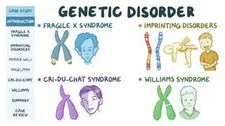
The four main types of structural chromosomal aberrations are deletion, duplication, inversion, and translocation. Deletions occur when a portion of the chromosome is deleted, or taken out, reducing or eliminating its function. For example, deletion of part of the short arm of chromosome 5 causes Cri-du-chat syndrome, characterized by reduced head size (microcephaly) and high-pitched crying in infants. In duplication, part of the chromosome is duplicated, resulting in extra genetic material. This occurs in Charcot-Marie-Tooth disease type I, in which part of chromosome 17 is duplicated, causing muscle weakness. Inversion of a chromosome happens when the genetic material is inverted, or flipped in the opposite direction. Inversions rarely cause disease and most commonly affect chromosome 2. Translocations occur when a piece of one chromosome has broken off from its original location and attaches to another chromosome. The most common example is a Robertsonian translocation, which occurs when two acrocentric chromosomes (chromosomes with arms of unequal lengths due to a non-centered centromere) lose their short arms, and the two long arms consequently join. Robertsonian translocations are a potential cause of trisomy. 

What are the most common chromosomal disorders?

Chromosomal disorders most commonly result from aneuploidy, most often trisomy. Notably, trisomy 21, or the presence of an extra chromosome 21, is the cause of Down syndrome. Down syndrome can present with a variety of signs and symptoms, including Brushfield spots, which are small white or grey spots on the edge of the iris, the colored part of the eye; dysmorphic features (e.g., atypically small head or flat facial profile); gastrointestinal problems; cardiovascular problems; neuromuscular problems (e.g., decreased muscle tone); pale skin; fatigue; and shortness of breath 

The other two most common autosomal trisomies are trisomy 18, which results in Edwards syndrome, and trisomy 13, which leads to Patau syndrome. Edwards syndrome often presents with severe mental disability, clenched hands, a large back of the head, a small mouth, low-set ears, and rocker-bottom feet, which are characterized by large heels. Signs of Patau syndrome include severe intellectual disability, a small head, small eyes, cleft lips or palate, more than five fingers on a hand (i.e., polydactyly), rocker-bottom feet, and malformation of the forebrain (i.e., holoprosencephaly). Advanced maternal age is a risk factor for all three trisomy disorders and for chromosomal aberrations in general. 

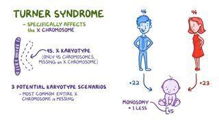
Aneuploidies of sex chromosomes can also occur, but they usually present with different symptoms than autosomal disorders. In general, signs and symptoms of sex chromosome aneuploidies include delayed puberty, absence of menstruation, ambiguous genitalia, and infertility. The most common sex chromosome disorder is Klinefelter syndrome, in which an individual with XY chromosomes has an additional X chromosome, or the genotype XXY, and is often assigned male at birth. The other two sex chromosome trisomies are XYY syndrome and XXX syndrome. Finally, Turner syndrome is the only sex chromosome monosomy, which occurs when a person inherits only one X chromosome and is usually assigned female at birth. 

What causes chromosomal aberrations?

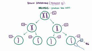
Chromosomal aberrations are most often caused by errors during cell division. Cell division in humans occurs via mitosis or, in sex cells, meiosis. In mitosis, cells duplicate their chromosomes and produce daughter cells with an identical number of chromosomes as the original cell. In other words, a cell with 46 chromosomes will produce two identical cells, each containing 46 chromosomes. Meanwhile, cell division by meiosis involves two rounds of cell division that allow for the recombination of genetic material, resulting in four unique sex cells, each with only half of the number of chromosomes. For example, a cell with 46 chromosomes undergoing meiosis will produce four unique daughter cells, each containing 23 chromosomes.  

Aneuploidy may result from an error during meiosis. Most commonly, this error results from nondisjunction  when a set of chromosomes does not properly separate - leaving one or two sex cells with an extra chromosome or missing one. If a sex cell affected by nondisjunction undergoes fertilization, the resulting offspring will inherit one extra or missing chromosome and may develop a chromosomal disorder 

Less commonly, aneuploidy can be the result of structural chromosomal aberrations. Structural chromosomal aberrations occur when part or all of a chromosome is missing, turned upside down, duplicated, or translocated to another chromosome. When this occurs after meiosis in a sex cell, it can lead to daughter cells with two copies or no copies of a chromosome, which may result in the offspring inheriting a monosomy or trisomy. 

Sometimes, chromosomal disorders are caused by mosaicism, a condition in which an individual has two or more different cell lines. Mosaicism can result from nondisjunction during mitotic cell division in early embryonic development. This produces one cell line with a chromosomal aberration while other lines remain unchanged.

How are chromosomal aberrations diagnosed?

Chromosomal aberrations in the offspring can often be detected through prenatal screening tests. A fetal ultrasound near the end of the first trimester or early in the second trimester can show increased nuchal translucency, which is a measure of the fluid-filled space in the back of the neck, potentially indicating trisomy 13, 18, or 21. A blood screening may also be conducted late in the first trimester to test for pregnancy-associated plasma protein A (PAPP-A) and human chorionic gonadotropin (hCG), which may indicate a chromosomal disorder if the levels are out of their expected ranges. During the second trimester, a “quad screen may be conducted on a blood sample, measuring levels of inhibin A, alpha-fetoprotein (AFP), hCG, and unconjugated estriol (uE3). A combination of high or low results can indicate different types of chromosomal disorders. Outside of the traditional maternal blood screenings, the same tests may be conducted on amniotic fluid obtained through amniocentesis (drawing of the fluid surrounding the fetus), or on placental tissue collected through chorionic villus sampling. 

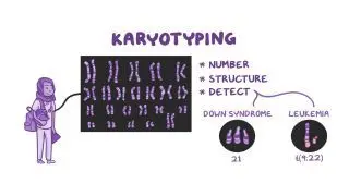
For diagnosis after birth, clinicians may assess the infant’s signs and symptoms and confirm the diagnosis using blood sample, including karyotyping and fluorescence in situ hybridization (FISH). Through karyotyping, all of the chromosomes are displayed visually in order to reveal any large structural abnormalities or numerical differences. Similarly, FISH uses fluorescent probes to visualize the genetic material, allowing detection of both structural and numerical abnormalities. 

How can you reduce the risk of chromosomal aberrations?

Parents may reduce the risk of chromosomal aberrations in their offspring by meeting their own nutritional needs, limiting exposure to problematic substances, and visiting a doctor before becoming pregnant. General risk reduction strategies include eating healthy, abstaining from smoking or drinking alcohol, and taking prenatal vitamins prior to pregnancy. Chromosomal aberrations are more likely to occur when the pregnant individual is over the age of 35. If a chromosomal disorder has been identified in a family, a healthcare provider may recommend genetic counseling to discuss different options, including assisted reproduction techniques. 

What are the most important facts to know about chromosomal aberrations?

Chromosomal aberrations are changes in chromosome structure or number. Most chromosomal aberrations are known as aneuploidies, or the presence of one extra or one missing chromosome within a pair. A trisomy is a type of aneuploidy with one extra chromosome, resulting in 3 copies, while a monosomy has one missing chromosome, leaving only a single copy. Structural chromosomal aberrations include any changes to a specific part of a chromosome, such as deletions, insertions, inversions, and translocations. The most common chromosomal disorder is trisomy 21, which results in Down syndrome. Some other chromosomal disorders include Edwards syndrome, Patau syndrome, Klinefelter syndrome, and Turner syndrome. Commonly, chromosomal aberrations occur due to problems during the division of sex cells inherited from a parent. Chromosomal aberrations may be diagnosed before or after birth through ultrasound imaging and laboratory tests. Parents may be able to reduce some risk of chromosomal aberrations by visiting a healthcare provider before pregnancy, having a nutritious diet, and avoiding smoking and alcohol consumption. 

Key Takeaways

Definition 
 

Chromosomal aberrations are changes in the structure or number of chromosomes.  

Types 
 

-Numerical = Aneuploidy  

-Monosomy 

-Trisomy 

-Structural 

-Deletion (Cri-du-chat syndrome) 

-Duplication (Charcot-Marie-Tooth disease type 1) 

-Inversion 

-Translocation (Robertsonian, often cause trisomies) 

Most Common 

-Autosomal chromosomes:  

-Trisomy 21 → Down syndrome 

-Trisomy 18Edward syndrome 

-Trisomy 13Patau syndrome  

-Sex chromosomes:  

-Trisomy XXY → Klinefelter syndrome 

-Monosomy X → Turner syndrome 

Causes 

-Nondisjunction or structural aberration during meiosis → extra or missing chromosome inherited by daughter sex cell  

-Mosaicism = nondisjunction error during mitosis in early embryonic development, involves only one cell line 

Diagnosis 

-Prenatal screening tests 

-Increased fetal translucency at ultrasound 

-Altered levels of PAPP-A and hCG 

-Quad screen 

-Can be done also on amniotic fluid or placental tissue 

-Postnatal diagnosis 

-Clinical signs and symptoms 

-Karyotyping 

-Fluorescent in situ hybridization (FISH) 

Risk Reduction 

-Prior to pregnancy:  

-Adequate diet and supplements  

-Avoid smoking and alcoholic beverages 

-Genetic counseling if a chromosomal disorder has been identified in a family  

-Increased risk for pregnant individuals older than 35 

Students say Osmosis is 100% worth it

Because Osmosis saves them time. Lowers stress. And actually helps them remember when it counts.

I used Osmosis to prepare for my first medical school licensing exam! Super helpful and interactive for people who may not do great with just pages of text info!

Cecilia Ruiz

Cecilia Ruiz

MD student

Sayan Misra

I have used Osmosis for about four years. Best thing I have ever used for my medical studies.

Sayan Misra

Sayan Misra

Med student

Osmosis videos are superior because they define simple concepts, tell a story with a clear progression, and provide context.

Jay Pate

Jay Pate

Dental student

References


Clancy S, Shaw K. DNA deletion and duplication and the associated genetic disorders. Nature Education. 2008;1(1):23. Retrieved from https://www.nature.com/scitable/topicpage/dna-deletion-and-duplication-and-the-associated-331/ 


Caceres V, Murray T, Myers C, Parbhoo K. Prenatal genetic testing and screening: a focused review. Semin Pediatr Neurol. 2022;42:100976. doi:10.1016/j.spen.2022.100976.Feuk L. Inversion variants in the human genome: role in disease and genome architecture. Genome Medicine. 2010;2(2):1-8. doi:10.1186/gm132 


Genetic Alliance; The New York-Mid-Atlantic Consortium for Genetic and Newborn Screening Services. Understanding Genetics: A New York, Mid-Atlantic Guide for Patients and Health Professionals. Washington (DC): Genetic Alliance; July 8, 2009. 


Nagamani SCS, Stankiewicz P, Lupski JR. GENE, genomic, 
and chromosomal disorders. In: Goldman L, Schafer AI, eds. Goldman-Cecil Medicine. 25th ed. Philadelphia, PA: Elsevier; 2016:chap 41.The heart & Down syndrome. In: National Down Syndrome Society, NDSS. Retrieved from https://www.ndss.org/resources/the-heart-down-syndrome/ (n.d.). 

Yilmaz F, Beck CR, Lee C. Disorders of the autosomes and sex chromosomes. In: Cohn R, Scherer S, Hamosh A, eds. Thompson & Thompson Genetics and Genomics in Medicine. 9th ed. Elsevier; 2023:79-108.