Corneal Dystrophy · What Is It, Diagnosis, Treatment, and More

Published: Apr 07, 2026
Author: Nikol Natalia Armata, MD
Editor: Alyssa Haag, MD
Editor: Ian Mannarino, MD, MBA
Editor: Kelsey LaFayette, DNP, ARNP, FNP-C
Illustrator: Abbey Richard, MSc
7-day free trial

Go deeper with Osmosis

Osmosis is a learning platform with videos, questions, and AI tools to help you master topics like this.

4.8 · 12,000+ reviews
Watch quick, visual videos
Practice with Qbank-style questions
Use AI to explain, quiz, and review
Study anytime with the mobile app
Start free trial

No credit card · Cancel anytime

What is corneal dystrophy?

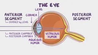
Corneal dystrophies (CD) refer to progressive conditions of the eye associated with the build-up of substances (e.g., proteins, lipids, amyloid) in one or more layers of the cornea. The cornea is the transparent front part of the eye, which consists of five distinct layers, each serving a crucial role in maintaining vision. The outermost layer of the cornea is the epithelium, acting as a protective barrier against external factors. Below the epithelium lies Bowman’s membrane, adding further resilience to the cornea. The stroma, the thickest and third layer, is primarily composed of water and collagen fibers, providing strength, refraction, and clarity while also allowing flexibility to the cornea. Descemet’s membrane, a thin but strong layer beneath the stroma, offers additional protection. Finally, the endothelium, the innermost layer, consists of cells responsible for regulating fluid balance within the cornea, ensuring optical clarity. The build-up of any substance in any of the above structures can lead to corneal dystrophy. 

Corneal dystrophies are grouped into three categories. Anterior or superficial corneal dystrophies, such as map-dot-fingerprint dystrophy, also called epithelial basement membrane dystrophy, affect the outermost layers of the cornea (i.e., the epithelium and Bowman’s membrane). Stromal corneal dystrophies, including lattice dystrophy, affect the stroma. Lastly, are posterior corneal dystrophies, which affect the innermost parts of the cornea (i.e., the endothelium and the Descemet membrane), such as Fuchs dystrophy.  

Learn deeper with Osmosis

Master this topic faster with videos, questions, and AI.

Used by 8M+ healthcare learners.

Start free trial

No credit card · Cancel anytime

What causes corneal dystrophy?

The causes of corneal dystrophies usually originate from genetic mutations affecting proteins involved in corneal structure and function. These mutations result in abnormal accumulation of lipids, proteins, or carbohydrates within the cornea, disrupting its transparency and smoothness. While the exact mechanisms underlying each type of corneal dystrophy may vary, inheritance patterns often follow autosomal dominance, meaning that only one copy of a mutated gene from either parent is sufficient to cause disease. There are autosomal recessive cases, in which two copies of a mutated gene, one from each parent, are required to cause some CDs as well. Some studies suggest that CD, particularly those with autosomal recessive inheritance patterns, can be more common in consanguineous families. 

Lisch epithelial CD and X-linked endothelial CD (XECD) are X-linked dominant diseases meaning that any mutation in just one copy of a gene on the X chromosome can cause CD. These X-linked disorders may be associated with other concomitantly inherited diseases, such as keratosis pilaris (i.e., benign condition causing dry, rough patches and tiny bumps on the skin), or congenital, inflammatory papules that result in hair loss. Environmental factors, such as UV exposure or eye trauma, may also influence disease progression in some cases. Additionally, certain corneal dystrophies, such as Fuchs dystrophy, may have multifactorial causes involving both genetic predisposition and other factors, such as aging, recurrent corneal erosion, or oxidative stress. 

What are the signs and symptoms of corneal dystrophy?

Individuals with CD may be asymptomatic or report vision problems, such as blurry vision, double vision, or refractive glares or halos. Symptomatic individuals may also complain of eye pain, with or without light sensitivity and foreign body sensation. These symptoms are due to corneal erosions, or the separation of the squamous epithelial layer from the anterior basement membrane. Pain typically occurs in the morning, after nocturnal drying of the cornea causing the epithelium to shear upon eyelid opening. Corneal erosions are common in almost all epithelial-based dystrophies. Generally, CD presents with visual alterations bilaterally, however, some dystrophies such as posterior polymorphous CD may present unilaterally. Another characteristic sign present in congenital stromal CD, is strabismus (i.e., the eyes do not properly align with each other when looking at an object) at birth. 

How is corneal dystrophy diagnosed?

Diagnosing corneal dystrophy entails a thorough examination conducted by an ophthalmologist, incorporating both medical history and specialized tests. Through slit-lamp examination, a detailed observation of the cornea under high magnification enables the detection of abnormal structures in the cornea. Additionally, corneal topography provides a detailed map of the corneal shape and reveals any curvature abnormalities. Optical coherence tomography (OCT) offers cross-sectional views, allowing the visualization and assessment of corneal layers. Specular microscopy, another valuable tool, examines corneal endothelial cells' density and morphology. In complex cases or when a genetic component is suspected, genetic testing may be employed to identify specific mutations associated with certain dystrophies, particularly when there is a family history of the condition or when the diagnosis is unclear based on clinical examination alone. 

How is corneal dystrophy treated?

Treatment for corneal dystrophy aims to alleviate symptoms, preserve vision, and address underlying causes whenever possible. Treatment plans for corneal dystrophy are individualized based on the type and severity of the condition, as well as the individual’s specific symptoms and needs. One common approach involves the use of medications, such as artificial tears and antibiotic eye drops or ointments, to manage symptoms like dryness, discomfort, or inflammation associated with the condition.  

Additionally, specialized soft contact lenses, such as rigid gas permeable or scleral lenses, may be prescribed to improve vision by masking irregularities in the corneal surface caused by dystrophy. In more advanced cases where vision loss is significant and other treatments prove ineffective, a corneal transplant (i.e., keratoplasty) may be necessary. This procedure involves replacing the damaged corneal tissue with healthy donor tissue to restore vision. 

Another treatment option is phototherapeutic keratectomy (PTK), a laser treatment that removes abnormal corneal tissue and smoothens the corneal surface. It can be beneficial for certain types of corneal dystrophies, particularly those affecting the outer layers of the cornea. Additionally, amniotic membrane transplantation may be utilized to promote corneal healing, reduce inflammation, and prevent scarring in some cases. 

Alongside medical interventions, symptomatic management through lifestyle modifications, such as wearing sunglasses to shield the eyes from UV radiation and avoiding eye rubbing, can also help alleviate symptoms and slow disease progression.

Emerging therapies are promising, with treatments such as enzyme therapy, gene therapy or RNA interference having positive results for targeting specific genetic forms of corneal dystrophy by addressing the defective enzymes or underlying genetic defects that lead to deposition of substances in the cornea 

What are the most important facts to know about corneal dystrophy?

Corneal dystrophy (CD) is a progressive condition affecting the eye caused by the accumulation of substances like proteins, lipids, and amyloid in the cornea affecting its transparency. CDs are categorized into anterior, stromal, and posterior types based on the affected corneal layer. They are usually genetic, with autosomal dominant, autosomal recessive, and X-linked inheritance patterns. Symptoms if present include vision problems, pain in the eye, light sensitivity and foreign body sensation. Diagnosis involves ophthalmologic exams and genetic testing, with treatments ranging from medication to corneal transplants. 

Key Takeaways

Definition 

Corneal dystrophies (CD) refer to progressive conditions of the eye associated with the build-up of substances (e.g., proteins, lipids, amyloid) in one or more layers of the cornea. 

The cornea 

-Transparent front later of the eye, 5 layers (from outermost):  

-Epithelium  

-Bowman's membrane  

-Stroma  

-Descemet's membrane  

-Endothelium  

Categories  

-Anterior or superficial corneal dystrophies  

-e.g., map-dot-fingerprint dystrophy (or epithelial basement membrane dystrophy)  

-Stromal corneal dystrophies  

-e.g., lattice dystrophy  

-Posterior corneal dystrophies  

-e.g., Fuchs dystrophy  

Causes 

-Genetic mutations → accumulation of abnormal proteins/lipids/carbohydrates within the cornea  

-Autosomal dominant, recessive, or X-linked 

-Environmental factors  

-UV exposure  

-Eye trauma  

-Often multifactorial (e.g., Fuchs dystrophy: genetic predisposition + aging + recurrent corneal erosion + oxidative stress 

Signs and symptoms 

-Asymptomatic  

-Vision problems:  

-Blurry vision  

-Double vision  

-Refractive glares or halos  

Strabismus (congenital stromal CD)  

-Eye pain (morning) 

-Light sensitivity  

-Foreign body sensation  

Diagnosis 

-Medical history  

-Slit-lamp examination  

-Corneal topography  

-Optical coherence tomography  

-Specular microscopy  

-Genetic testing  

Treatment 

-Symptoms management:  

-Artificial tears  

-Antibiotic eye drops/ointment  

-Specialized soft contact lenses  

-Lifestyle modification (sunglasses, avoid eye rubbing) 

-If significant vision loss: corneal transplant  

-In selected cases: phototherapeutic keratectomy (PTK), amniotic membrane transplantation  

-Emerging treatments: enzyme therapy, gene therapy, RNA interference 

Students say Osmosis is 100% worth it

Because Osmosis saves them time. Lowers stress. And actually helps them remember when it counts.

I used Osmosis to prepare for my first medical school licensing exam! Super helpful and interactive for people who may not do great with just pages of text info!

Cecilia Ruiz

Cecilia Ruiz

MD student

Sayan Misra

I have used Osmosis for about four years. Best thing I have ever used for my medical studies.

Sayan Misra

Sayan Misra

Med student

Osmosis videos are superior because they define simple concepts, tell a story with a clear progression, and provide context.

Jay Pate

Jay Pate

Dental student

References


Klintworth GK. Corneal dystrophies. Orphanet J Rare Dis. 2009;4:7. Published 2009 Feb 23. doi:10.1186/1750-1172-4-7 


Lisch W, Weiss JS. Clinical and genetic update of corneal dystrophies [published correction appears in Exp Eye Res. 2020 Jan 24;:107929]. Exp Eye Res. 2019;186:107715. doi:10.1016/j.exer.2019.107715 


Salman M, Verma A, Singh VK, et al. New frontier in the management of corneal dystrophies: Basics, development, and challenges in corneal gene therapy and gene editing. Asia Pac J Ophthalmol (Phila). 2022;11(4):346-359. doi:10.1097/APO.0000000000000443 


Soh YQ, Kocaba V, Weiss JS, et al. Corneal dystrophies. Nat Rev Dis Primers. 2020;6(1):46. Published 2020 Jun 11. doi:10.1038/s41572-020-0178-9