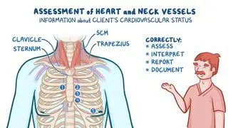
To diagnose constrictive pericarditis, a clinician will often first assess the individual’s vital signs, symptoms, and medical history. One of the primary signs of constrictive pericarditis is hearing a pericardial knock during diastole when a provider listens to the heart sounds with a stethoscope. In addition, constrictive pericarditis is often accompanied by progressive shortness of breath, fatigue, and weakness. People with constrictive pericarditis usually present with chronic swelling of legs and arms (i.e., edema) and abdominal swelling (i.e., ascites). Another indicator of constrictive pericarditis is the Kussmaul sign, which can be observed when the individual’s jugular vein, the large vein in the neck, sticks out when the individual breathes in.
Afterwards, diagnostic imaging may be performed, most commonly electrocardiography and echocardiography. An electrocardiography (ECG) records electrical impulses from the heart and may show a nonspecific finding, known as diffuse ST-T wave changes. In severe cases, the ECG can show an irregular, fast heart beat, known as atrial fibrillation. Unlike ECG, echocardiography creates images by using sound waves, and two types may be used for diagnosis of constrictive pericarditis: 2-D or Doppler. 2-D echocardiography can help visualize the thickened pericardium and other changes to the heart. Doppler echocardiography may show an unusually rapid early diastolic filling.
Other tests, such as cardiac catheterization, in which a thin tube is inserted into the heart, or cardiac magnetic resonance imaging (MRI) may be necessary if diagnosis remains unclear. Occasionally, a chest X-ray may also be performed.
Without treatment, constrictive pericarditis can lead to
heart failure and become life threatening. Initial treatment for constrictive pericarditis focuses on treating the underlying cause. If inflammation continues,
anti-inflammatory medications or
diuretics may be prescribed, and individuals may be recommended to reduce salt intake. However, constrictive pericarditis is often permanent and progressive, usually requiring a pericardiectomy, in which a portion or all of the pericardium is surgically removed from the heart. This surgery can be dangerous, and as the
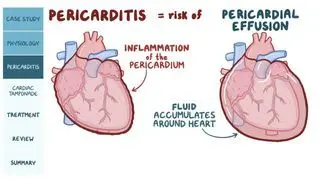
pericarditis progresses, the risk of complication increases.