Arrhythmias - Asystole: Nursing
Introduction0:00–0:39
Cardiac arrest occurs when the heart suddenly stops pumping blood throughout the body. There are two types of cardiac arrest, asystole or cardiac flatline, and pulseless electrical activity, or PEA for short.
Asystole is the most common type and refers to the total absence of electrical and mechanical activity of the heart. On the other hand, PEA occurs when there is electrical activity in the heart, but the cardiac muscle is unresponsive to stimulation; therefore, there’s no mechanical activity of the heart and pulse.Now, let’s cover some physiology of the cardiac conduction system.
Physiology0:39–2:59
The cardiac conduction system is made up of specialized myocardial cells that can create and transport electrical potential, also called an action potential.
These cells have many special features, including automaticity, meaning that they can generate an impulse, excitability, which is the ability to respond to a stimulus by creating an electrical impulse, conductivity meaning they can carry the impulse to other cells, and contractility, which is the ability to shorten the length of their fibers, causing a contraction.
Now let’s look at the normal electrical conduction pathway in the heart on an ECG, which shows how the depolarization wave flows through the heart during each heartbeat.
The normal electrical activity of the heart starts in the sinoatrial node or SA node, which is considered the pacemaker of the heart.
Then, the impulse is conducted through the atrium, creating the P wave on an ECG. And when the atrial muscle cells get depolarized, they contract, pushing blood from the atria into the ventricles.
From the atrium, electrical activity goes to the atrioventricular node, or AV node, where the impulse propagation speed slows way down; this is the PR interval on an ECG.
This pause allows the atria to contract while the ventricles fill with blood. From the AV node, the depolarization wave goes through the Bundle of His, then the right and left branches of the Bundle, and finally through the Purkinje fibers, which deliver the current to the right and left ventricles, causing them to depolarize.
This triggers simultaneous contraction of both ventricles, pushing blood into the systemic and pulmonary circulations, and it’s represented by the QRS complex on an ECG.
Finally, the ventricles repolarize to prepare for the next cycle, which allows them to relax and fill with blood, called diastole.
And on ECG, ventricular repolarization will create a T wave, while the pause between ventricular depolarization and repolarization is represented by the ST segment.
Sometimes, immediately after the T wave, there’s a U wave, which represents late repolarization of the ventricles. Now, based on the cause, asystole can be classified as primary and secondary.
Causes & risk factors2:59–4:18
Primary asystole occurs when the problem is at the level of the cardiac conduction system. On the other hand, secondary asystole occurs when some other condition that is not related to the cardiac conduction system, affects the heart’s ability to fire and conduct electrical impulses.
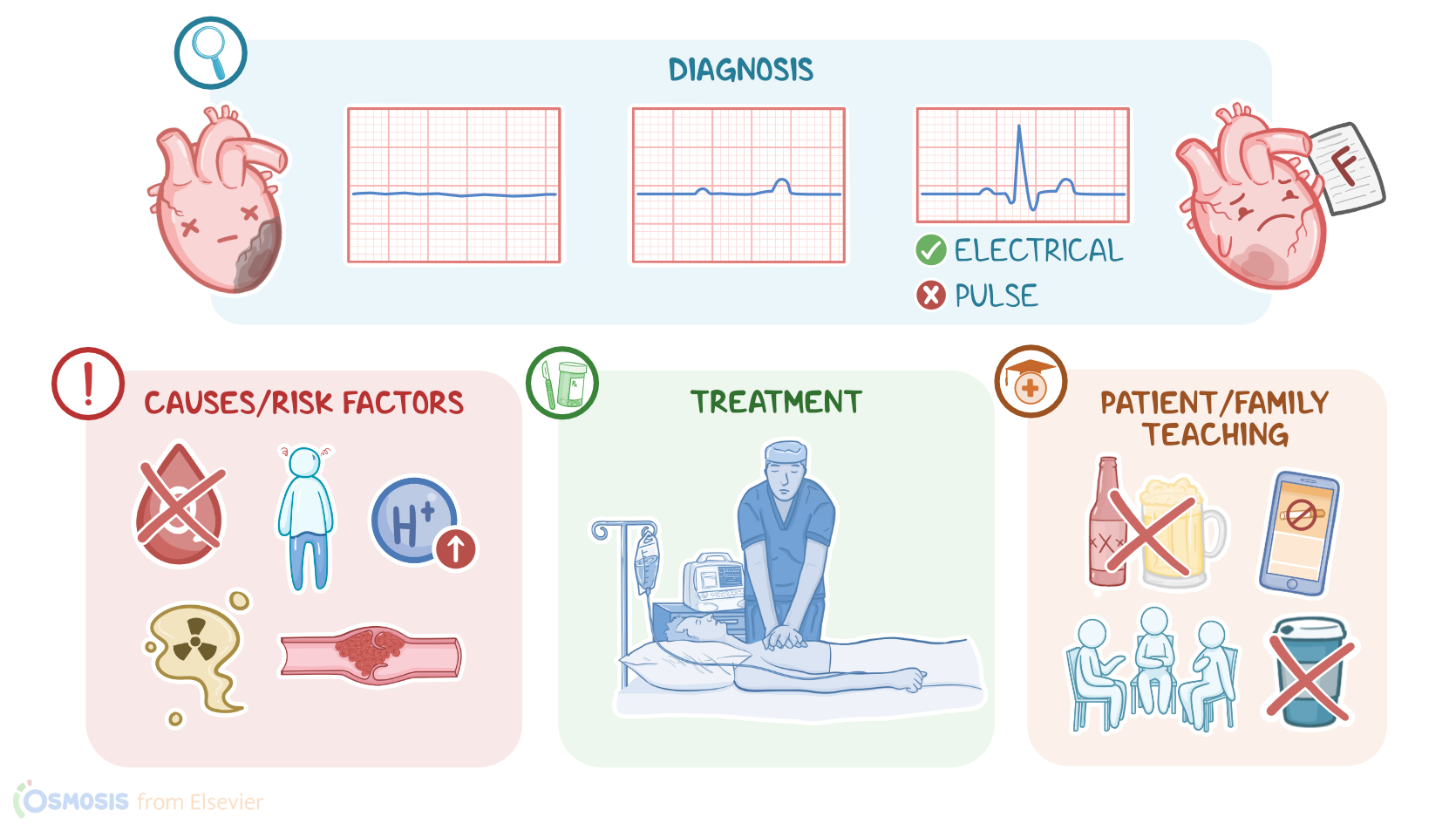
Secondary causes can be further subdivided into two main groups: 5 H’s and 5 T’s. 5 Hs include Hypoxia, or low levels of oxygen in peripheral tissues; Hypovolemia, or volume depletion; Hypothermia, or low body temperature; high concentration of Hydrogen ions, or acidosis; and finally, Hypo- or Hyperkalemia.
On the flip side, 5 Ts include Toxins, cardiac Tamponade, Tension pneumothorax, pulmonary Thrombosis, and coronary Thrombosis.
Important risk factors associated with asystole include medical history of cardiovascular conditions, ventricular fibrillation or V-fib, and ventricular tachycardia or V-tach.
Additionally, clients who are assigned female at birth, and children are more likely to develop asystole.Now, moving on to pathology.
Asystole typically occurs due to an abnormal ventricular rhythm, like V-fib or V-tach, which causes cessation of the electrical activity.
Pathology4:18–4:50
As a result, ventricles fail to depolarize, contract, and pump blood throughout the body. When there’s not enough blood circulating through the body, there’s no oxygen for peripheral tissues, so they are left deprived.
This can result in severe complications, like permanent brain damage, brain death, or even full body death. Clinical manifestations of asystole include sudden collapse with loss of consciousness, unresponsiveness, and apnea, meaning cessation of breathing.
Clinical manifestations4:50–5:08
Upon physical assessment, the client is usually pulseless with absent heart sounds and no recordable blood pressure.Diagnosis of asystole is based on the client’s history, physical assessment, and electrocardiographic features, which include isoelectric flatline and the absence of ventricular activity.
Diagnosis5:08–5:34
Other clients might have P waves but without any QRS complexes. It’s important to note that clients with PEA have normal ECG features, although their heart is not pumping, so physical assessment is crucial.
Treatment of asystole consists of cardiopulmonary resuscitation and intravenous epinephrine. Clients with secondary asystole also require the treatment of the underlying cause.
Treatment5:34–6:08
Finally, keep in mind that asystole and PEA are non-shockable heart rhythms, which means that defibrillation cannot be used to correct asystole, because there is no electrical activity to “reset”, or PEA because the cardiac muscle is unresponsive to electrical stimulation.
Alright, let’s look at the nursing care you’ll be providing for a client in asystole. Your priority nursing goal is to restore a perfusing heart rhythm.Okay, if your client is in asystole, call for assistance and begin chest compressions, as other team members administer 100% oxygen, provide bag-mask ventilation, and administer epinephrine, per protocol.
Management and care6:08–7:07
Continue with CPR as the team intubates your client, and identifies and treats the underlying cause, as indicated. Continue to assess for signs of return of spontaneous circulation, or ROSC, including the presence of a pulse, a measurable blood pressure, or an increase in end-tidal CO2.
Once ROSC is achieved, provide post-cardiac arrest care and coordinate transfer to ICU for hemodynamic stabilization and recovery.Now, moving on to client and family teaching.
General client and family teaching7:07–8:18
Begin by explaining that asystole occurs when the heart suddenly stops pumping blood throughout the body.Next, talk to your client about lifestyle modifications that can help keep their heart healthy.
Encourage them to maintain a healthy weight, and to engage in moderate physical exercise regularly, as prescribed by their healthcare provider.
Instruct them to eat plenty of fruits, vegetables, and whole grains, as well as lean meats, and low-fat or fat-free dairy products.
If they need help quitting smoking, provide them with counseling and refer them to resources for support.Also, teach them how to check their pulse and blood pressure, and encourage family members to learn CPR.
Instruct them to seek emergency care if they experience symptoms like chest pain, difficulty breathing, dizziness, or fainting.All right, as a quick recap.
Review8:18–9:07
Asystole refers to any event that leads to cardiac arrest. Possible causes that lead to cardiac arrest are: hypoxia, hypovolemia, hypothermia, high concentration of hydrogen ions or acidosis, hypo- or hyperkalemia; toxins, cardiac tamponade, tension pneumothorax, pulmonary thrombosis, or coronary thrombosis.
Treatment of asystole consists of high quality CPR as recommended by AHA/BLS guidelines. Recovery of the client once ROSC is established continues in ICU for hemodynamic stability.
The priority goal of nursing care is to restore a perfusing heart rhythm. Client and family teaching is focused on maintaining a healthy lifestyle and when to seek emergency care.
| ARRHYTHMIAS - ASYSTOLE | ||
| KEY POINTS | NOTES | |
| DEFINITION |
| |
| PHYSIOLOGY |
| |
| CAUSES AND RISK FACTORS |
| |
| PATHOPHYSIOLOGY |
| |
| SIGNS AND SYMPTOMS |
| |
| DIAGNOSIS |
| |
| TREATMENT |
| |
| MANAGEMENT OF CARE |
| |
| PATIENT AND FAMILY TEACHING |
| |
- "Critical care nursing: Diagnosis and management (10th ed.) ISBN: 978-0-443-11581-3 " Elsevier (2026)
- "Lewis’s Medical-Surgical Nursing: Assessment and Management of Clinical Problems. 12th Edition. ISBN:978-0-323-78961-5 " Elsevier
- "Mosby’s® Diagnostic and Laboratory Test Reference. 15th edition. ISBN: 978-0-323-67519-2 " Mosby (2020)
- "Saunders Comprehensive Review for the NCLEX-RN. 9th Edition. ISBN: 978-0-323-79530-2 " Saunders (2022)
- "Fundamentals of Nursing. 11th edition. ISBN: 978-0-323-81034-0 " Elsevier (2022)
- "Seidel’s Guide to Physical Examination: An Interprofessional Approach. 10th edition. ISBN: 978-0-323-76183-3 " Elsevier (2022)
- "Critical Care Nursing: Diagnosis and Management. 9th edition. ISBN: 978-0-323-64295-8 " Elsevier (2021)
- "Invasive Management of Acute Myocardial Infarction Complicated by Cardiogenic Shock: A Scientific Statement From the American Heart Association. 143(15):e815-e829." Circulation (2021)
- "Obesity and Cardiovascular Disease: A Scientific Statement From the American Heart Association. 143(21):e984-e1010." Circulation (2021)
- "Best Practices for Education and Training of Resuscitation Teams for In-Hospital Cardiac Arrest. 14(12):e008587." Circ Cardiovasc Qual Outcomes. (2021)
No notes for this video yet
Try adding a note below