Arrhythmias - Ventricular tachycardia (Vtach): Nursing
Introduction0:00–0:52
Arrhythmias are irregular heartbeats that occur due to any disturbance in the rate, rhythm, site of origin, or conduction of the cardiac electrical impulse.
Which can affect the heart's ability to pump blood throughout the body. Ventricular tachycardia, or simply VTA, is a type of tachyarrhythmia, where a point in the ventricles fires abnormal signals, causing the heart to beat faster than normal, at a rate of 150 to 250 BPM.
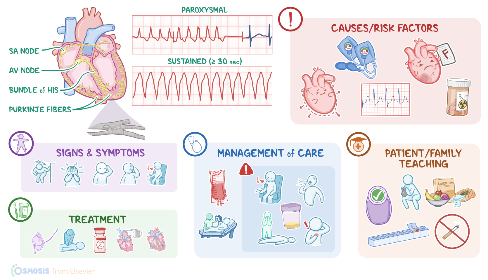
Ventricular tachycardia can be paroxysmal, meaning that it presents as 3 or more beats that are self-limiting, or sustained, meaning that it lasts at least 30 seconds.
Now, the cardiac conduction system is made up of specialized myocardial cells that can create and transport electrical potential, also called an action potential.
Physiology0:52–2:38
These cells have many special features, including automaticity, meaning they can generate an impulse, excitability, which is the ability to respond to a stimulus by creating an electrical impulse, conductivity, meaning they can carry the impulse to other cells, and contractility, which is the ability to shorten the length of their fibers, causing a contraction.
All right, now, let's look at the normal electrical conduction pathway in the heart on an ECG which shows how the depolarization wave flows through the heart during each heartbeat.
The normal electrical activity of the heart starts in the sinoatrial or SA node, which is considered the pacemaker of the heart.
Then, the impulse is conducted through the atria, causing depolarization, and creating the P wave on an ECG. When the atrial muscle cells get depolarized, they contract, pushing blood from the atria into the ventricles.
From the atria, the impulse goes to the atrioventricular or AV node, where the impulse propagation speed slows way down.
The interval from the atrial depolarization to just before ventricular depolarization is the PR interval on an ECG. This delay allows the atria to contract, while the ventricles fill with blood.
From the AV node, the impulse goes through the bundle of hiss, then to the right and left bundle branches, and finally, through the perkinji fibers, which deliver the impulse to the right and left ventricles, causing them to depolarize, and is represented by the QRS complex on an ECG.
This triggers simultaneous contraction of both ventricles, pushing blood into the systemic and pulmonary circulations. Finally, the ventricles re-polarize to prepare for the next cycle, which allows them to relax and fill with blood, called diastole.
Causes & risk factors2:38–3:38
And on ECG ventricular repolarization will create a T wave, while the phase between ventricular depolarization and repolarization is represented by the ST segment.
Sometimes, immediately after the T wave, there's a U wave, which represents late repolarization of the ventricles. OK, now back to ventricular tachycardia, which is typically caused by any kind of structural damage to the heart that leads to electrical changes in the heart muscle cells.
These changes are strongly associated with several risk factors, particularly certain heart conditions. These include ischemic heart disease, previous or recent myocardial infarction, in addition to cardiomyopathy, supraventricular tachycardia, valvular heart disease, and heart failure.
Pathology3:38–5:10
In addition, clients with electrolyte imbalances, such as hypokalemia and hypomagnessemia, as well as chronic obstructive pulmonary disease or COPD are at an increased risk for ventricular tachycardia.
Finally, other risk factors include drug toxicity, typically with steroids, in addition to cocaine and use of inhalants.
Now, the pathology of ventricular tachycardia starts when the ventricular muscle cells develop ectopic nodal automaticity, or re-entrance signaling.
Eectopic nodal automaticity suggests that a ventricular cell called a ventricular ectopic focus, starts firing at higher rates that exceed the rate of the firing of the SA node, which increases the heart rate to more than 150 BPM.
What messes things up, is that the SA node may continue to send off signals, causing contraction of the atria that is independent of the contraction of the ventricles.
On the other hand, re-entrance signaling happens when a depolarization wave encounters tissue that doesn't depolarize, which could be due to the presence of scar tissue after myocardial infarction.
Here, the wave starts going around and around that tissue, forming what's called a re-entrant loop. Ultimately, these mechanisms cause irritability of ventricular cells, which leads to ventricular tachycardia.
Clinical manifestations5:10–5:33
When it comes to complications of ventricular tachycardia, these include hypotension, pulmonary edema, decreased cerebral blood flow, as well as an increased risk of developing ventricular fibrillation, which can lead to a sudden cardiopulmonary arrest or sudden death.
Clients with ventricular tachycardia typically present with dizziness, altered mental status, pallor, as well as diaphoresis and hypotension.
Diagnosis5:33–7:11
Clinical manifestations can also include chest pain, palpitations, and shortness of breath. Additionally, clients with ventricular tachycardia may or may not have a pulse.
The diagnosis of ventricular tachycardia starts with the client's history and physical assessment, followed by electrocardiography, which may show particular changes related to ventricular tachycardia.
These changes include a ventricular rate of 150 to 250 BPM, with a regular or irregular rhythm. Typically, clients with ventricular tachycardia have their P waves buried within the QRS complex, but in some cases, AV dissociation might be present, which means that the P waves occur independently from the QRS complex.
Additionally, the PR interval cannot be measured, and the T wave is in the opposite direction of the QRS complex, which is typically wide, and lasts for more than 0.12 seconds.
The QRS complex can also be distorted, meaning that it can have different shapes, and these shapes can be used to group ventricular tachycardia into three categories, monomorphic, polymorphic, and torso de point.
In monomorphic ventricular tachycardia, the QRS complexes have the same size, shape, and direction, whereas in polymorphic ventricular tachycardia, the QRS complex keeps changing from one size, shape and direction to another, over a set of beats.
Treatment7:11–8:59
However, in torsa de Point, which is French for twisting of the points, there's a prolongation of the QT interval, and the QRS complexes twist around the isoelectric line.
Alright, now the treatment of ventricular tachycardia starts with continuous ECG monitoring, and supplemental oxygen. Next, it's crucial to quickly determine if the client is hemodynamically stable, meaning they can maintain adequate BP and cardiac output.
Clinically unstable clients who are pulseless or unresponsive are considered to be in cardiac arrest. So, the first step is cardiopulmonary resuscitation, or CPR, defibrillation, and epinephrine.
For unstable clients who are not in cardiac arrest, treatment includes electrical cardioversion with a defibrillator and anti-arrhythmic medications.
When the client is hemodynamically stable, the treatment for ventricular tachycardia involves these same IV anti-arrhythmics.
Next, it's important to look for and address the underlying cause of the ventricular tachycardia, such as electrolyte imbalance or medications.
For long-term management, beta-blockers are typically given to help prevent future ventricular tachycardias. If they have other heart conditions like a previous myocardial infarction or heart failure, they may need an implantable cardiac defibrillator, or ICD.
However, ICDs can only stop ventricular tachycardias from progressing. So if the client still has multiple episodes, it can be treated with an oral anti-arrhythmic or radiofrequency ablation, where a catheter emitting high-frequency energy is used to destroy the small section of heart tissue that is causing the arrhythmia.
Management and care8:59–10:34
All right, let's look at the nursing care you'll provide to a client with ventricular tachycardia. Your priority goal is to assist in restoring a normal sinus rhythm.
OK, act quickly to work with the team to restore normal sinus rhythm, since ventricular tachycardia can be life-threatening.
First, determine if your client is hemodynamically stable by assessing vital signs and checking for symptoms of decreased cardiac output.
Report decreased LOC, hypotension, weak or absent pulse, chest pain, tachypnea, shortness of breath, or decreased urinary output to the healthcare provider.
Administer supplemental oxygen and prepare to assist with emergent synchronized cardioversion. Now, if your client is hemodynamically stable, administer the prescribed IV fluids and anti-arrhythmic medications.
Then, review their latest laboratory test results to help identify the underlying cause, including an electrolyte panel, cardiac enzymes, and toxicology screen.
Continue to monitor your client's ECG and hemodynamic status, and immediately report if your client becomes unresponsive or pulseless.
Initiate cardiopulmonary resuscitation for cardiac arrest. When spontaneous return to circulation returns, provide post-cardiac arrest care.
Lastly, administer treatments to address the underlying cause as prescribed. OK, let's move on to client and family teaching.
General client and family teaching10:34–12:16
First, explain to your client that ventricular tachycardia is a rapid heart rate that is caused by abnormal firing of signals in the ventricles of the heart.
Then, review the treatments that used to help the heart rhythm return to normal, and go over each of their prescribed medications, emphasizing the need to take them exactly as prescribed.
Next, talk to your client about lifestyle modifications that can help keep their heart healthy. Encourage them to maintain a healthy weight, and to engage in moderate physical exercise regularly, as prescribed by their healthcare provider.
Instruct them to eat plenty of fruits and vegetables, and whole grains, as well as lean meats, and low-fat or fat-free dairy products.
If they need help quitting smoking, provide them with counseling and refer them to resources for support. Also, teach them how to check their pulse and BP, and encourage family members to learn CPR.
Instruct them to notify their healthcare provider immediately if their heart rate is 100 BPM or more, or if they experience palpitations.
Also, urge them to seek emergency care if they experience symptoms like chest pain, difficulty breathing, dizziness, or fainting.
Alright, it's a quick recap. Ventricular tachycardia is a tachyarrhythmia caused by an abnormal electrical signal firing in the ventricles that leads to a dangerously rapid heart rate of 150 to 250 BPM.
Review12:16–14:25
Paroxysmal ventricular tachycardia is self-limiting, whereas sustained ventricular tachycardia lasts for 30 seconds or more.
Risk factors include ischemic heart disease, history of myocardial infarction, cardiomyopathy, supraventricular tachycardia, valvular heart disease, heart failure, electrolyte imbalances, COPD and drug toxicity.
These risk factors can lead to ectopic nodal automaticity, or re-entrance signaling, which ultimately results in ventricular tachycardia.
Complications to look out for include hypotension, pulmonary edema, decreased cerebral blood flow, and ventricular fibrillation, which can lead to sudden cardiopulmonary arrest, or sudden death.
Clinical manifestations with which clients typically present include dizziness, altered mental status, pallor, diaphoresis, hypotension, chest pain, palpitations, shortness of breath, and they may or may not have a pulse.
Diagnosis involves the client's history and physical assessment, as well as electrocardiography, which is used to classify ventricular tachycardia into being monomorphic, polymorphic, or torso depo.
Treatment is focused on identifying and treating the underlying cause, and depends on the type of ventricular tachycardia.
As well as whether the client is hemodynamically stable. Based on these factors, treatment may include anti-arrhythmics, cardioversion, and CPR along with defibrillation.
The priority goal of nursing care is to assist in restoring normal sinus rhythm. Finally, client and family education centers around health maintenance measures and when to seek medical attention.
| ARRHYTHMIAS - VENTRICULAR TACHYCARDIA (VTACH) | ||
| KEY POINTS | NOTES | |
| DEFINITION |
| |
| PHYSIOLOGY |
| |
| CAUSES AND RISK FACTORS |
| |
| PATHOPHYSIOLOGY |
| |
| SIGNS AND SYMPTOMS |
| |
| DIAGNOSIS |
| |
| TREATMENT |
| |
| MANAGEMENT OF CARE |
| |
| PATIENT AND FAMILY TEACHING |
| |
No notes for this video yet
Try adding a note below