Atelectasis: Nursing
Introduction0:00–0:14
Atelectasis is a condition where the alveoli in a lung subsegment or the entire lung collapse, inhibiting gas exchange.Now, let’s quickly review the lower respiratory tract, which includes the lower part of the trachea, and the lungs containing the bronchi, bronchioles, alveolar ducts, and finally the alveoli.
Physiology0:14–1:02
Alveoli are tiny air-filled sacs where most gas exchange occurs, so as we breathe, the inhaled oxygen moves from the alveolar sacs into the blood, while the carbon dioxide moves from the blood into the alveolar sacs to be exhaled.
The walls of alveoli are coated in surfactant, an oily secretion that reduces surface tension at the alveolar air interface and prevents the walls of the alveoli from sticking to each other.Alright, now the causes of atelectasis can be classified as obstructive and non-obstructive.
Causes & risk factors1:02–2:39
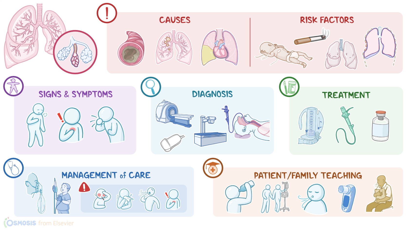
Obstructive atelectasis is the most common and results from obstruction of the bronchi between the alveoli and the trachea.
This prevents gas from moving into the alveoli, so they collapse. Causes of obstructive atelectasis include foreign objects, tumors, retained secretions and mucus plugs.
In non-obstructive atelectasis, there’s something pressing on the lungs, which results in the alveoli being physically compressed.
Some causes can include large pleural effusions and chest trauma. Non-obstructive atelectasis can also be caused by the lack of surfactant, which might be encountered in premature newborns.
Now, risk factors for atelectasis include premature birth, lifestyle factors, such as smoking and obesity, lung diseases like COPD, asthma, cystic fibrosis, and bacterial infections.
Other risk factors include anything leading to hypoventilation like spinal cord injuries, sedation from recent surgery, especially with general anesthesia; as well as pain, and prolonged immobility.
Lastly, there’s increased risk of developing atelectasis for clients who have chest wall abnormalities, such as scoliosis, rib fractures, or other trauma.Alright, now in atelectasis, the collapsed alveoli cannot participate in gas exchange causing reduced lung volume, and shunting, where blood doesn't become oxygenated as it moves through the lungs.
Pathology2:39–3:01
If the affected lung area is large enough, this can ultimately result in hypoxemia.Now, the clinical manifestations of atelectasis vary depending on the extent of alveolar collapse.
Clinical manifestations3:01–3:56
If only a small area of atelectasis is present, a client might be asymptomatic. If a larger area is affected, a client may experience shortness of breath, cough, chest pain, and respiratory distress, which presents as short, shallow, and rapid breathing, as well as decreased SpO2.During auscultation, there are reduced or absent breath sounds on the affected side.
Also, crackles can sometimes be heard, which is the sound of collapsed alveoli popping open with inspiration. On percussion, there’s dullness over the affected area, and on palpation, there might be decreased fremitus.Diagnosis of atelectasis begins with the client’s history and physical assessment.
Diagnosis3:56–4:17
A chest X-ray can show reduced lung volume. Finding the underlying cause of atelectasis may require additional testing such as a chest CT, bronchoscopy, or thoracic ultrasound.
Treatment of atelectasis generally focuses on re-expanding the collapsed alveoli and removing retained secretions. So, most clients receive chest physiotherapy which uses a variety of techniques to help clear the airway including encouraging coughing, deep breathing exercises, using an incentive spirometer, chest percussion, and postural drainage.
Treatment4:17–5:45
A bronchoscopy can be performed to remove obstructions such as foreign bodies, or mucus plugs. Mucolytic medications such as acetylcysteine may also be prescribed to break down mucus.
Mechanical ventilation like continuous positive airway pressure can be also used, which delivers pressurized air or oxygen through a nose or face mask to help ensure that the alveoli do not collapse.
In addition, clients with mechanical ventilation or tracheostomy may require suctioning to help them remove thick secretions like mucus from their airways.
Clients with complications like large effusions, hemothorax, or empyema require pleural drainage. Finally, depending on the underlying condition, clients might require surgical tumor resection, as well as radiation and chemotherapy or a thoracentesis for fluid build-up.Alright, let’s look at the nursing care you’ll provide for a client with atelectasis.
Management and care5:45–7:11
Your priority nursing goals are to promote lung expansion and prevent complications. Begin by assessing your client's vital signs, including respiratory rate, respiratory effort, lung sounds, and oxygen saturation.
Be sure to position your client with the head of the bed raised at least 30 degrees in order to maximize diaphragmatic excursion and ventilation, and help them with deep breathing and use of an incentive spirometer.
Manage secretions by providing chest physiotherapy, suctioning, and maintaining adequate hydration. Be sure to regularly reposition them every 1 to 2 hours, as needed, and assist them with ambulation.
Also administer the prescribed analgesics, as well as supplemental oxygen or mucolytics if indicated. Immediately report if your client develops signs or symptoms of pneumonia, such as fever, chills, productive cough, tachypnea, or pleuritic chest pain.
Be prepared to provide supplemental oxygen, obtain a sputum sample for culture, and administer the prescribed analgesics and antibiotics, as ordered.Now, moving on to client and family teaching.
General client and family teaching7:11–8:40
Explain what atelectasis is and how it can happen when they are hospitalized, especially after a surgical procedure; and let them know how promoting lung expansion and mobilizing secretions can help treat the atelectasis and prevent it from getting worse.
Also teach your client to take in plenty of fluids, which helps prevent the mucus in their lungs from thickening and forming plugs that can block the airways.
Next, let them know you will assist them to change their position in bed every 2 hours, and that you’ll be helping them with ambulation as soon as it’s safe to do so.
Then, show them how to use their incentive spirometer, and instruct them to use it every hour when they are awake. Reassure them that you’ll be working with them to control their pain so they can perform these tasks to help clear the atelectasis from their lungs.
Lastly, instruct them to call you or another healthcare provider right away if they experience chills, dyspnea, increased fatigue, if they feel like their cough is getting worse, or if their pain isn’t being controlled adequately.
Alright, as a quick recap… Atelectasis is a condition in which the alveoli in a lung section or the entire lung collapse, leading to impaired gas exchange.
Review8:40–10:48
Causes of atelectasis can be classified as obstructive, such as mucus plugs or tumors; and non-obstructive, due to pressure from outside the lungs, such as pleural effusion, chest trauma, or from lack of surfactant.
Risk factors for atelectasis include premature birth; lifestyle factors like smoking or obesity; lung diseases; chest wall abnormalities; and anything leading to hypoventilation, such as spinal cord injuries, sedation and anesthesia from recent surgery, pain, and prolonged immobility.
Now, the collapsed alveoli cannot participate in gas exchange, so blood doesn't become oxygenated as it moves through that part of the lungs.
If the affected lung area is large enough, this can ultimately result in hypoxemia. Clinical manifestations include shortness of breath, cough, chest pain, and respiratory distress; as well as crackles, reduced or absent breath sounds on the affected side upon auscultation; and dullness on percussion over the affected side.
Diagnosis of atelectasis includes a thorough history and physical assessment, followed by a chest X-ray, which shows reduced lung volume.
Additional testing such as a chest CT scan, bronchoscopy, or thoracic ultrasound may help diagnose atelectasis. Treatment focuses on re-expanding the collapsed alveoli and removing retained secretions in order to promote adequate gas exchange.
Nursing goals include promoting lung expansion and preventing complications. Client and family education is focused on measures they can take to restore lung volume and promote oxygenation.
| ATELECTASIS | ||
| KEY POINTS | NOTES | |
| DEFINITION |
| |
| PHYSIOLOGY |
| |
| CAUSES AND RISK FACTORS |
| |
| PATHOPHYSIOLOGY |
| |
| SIGNS AND SYMPTOMS |
| |
| DIAGNOSIS |
| |
| TREATMENT |
| |
| MANAGEMENT OF CARE |
| |
| PATIENT AND FAMILY TEACHING |
| |
No notes for this video yet
Try adding a note below