Intracranial aneurysm: Nursing
Intracranial aneurysm: Nursing
Watch later
Watch later
Notes
| INTRACRANIAL ANEURYSM | ||
| KEY POINTS | NOTES | |
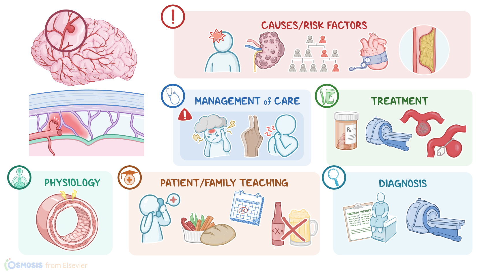
| DEFINITION |
| |
| PHYSIOLOGY |
| |
| CAUSES AND RISK FACTORS |
| |
| PATHOPHYSIOLOGY |
| |
| SIGNS AND SYMPTOMS |
| |
| DIAGNOSIS |
| |
| TREATMENT |
| |
| MANAGEMENT OF CARE |
| |
| PATIENT AND FAMILY TEACHING |
| |
Transcript
Aneurysms are abnormal dilations in a blood vessel that form in weakened areas of the blood vessel walls. Aneurysms can happen to any blood vessel in your body, including the cerebral vessels, in which case they’re called intracranial or cerebral aneurysms.
Alright, let’s go over some physiology. There are three major types of blood vessels: arteries, veins, and capillaries. Normally, blood flows from large arteries into medium and then small arteries called arterioles, which in turn carry the blood to capillary beds.
All arterial vessels have three layers: from outside in, there’s tunica externa or the adventitia layer, which has loose connective tissue, and sometimes, vasa vasorum or vessels that supply the artery; then tunica media or the media layer, which contains some elastic tissue and smooth muscle that allow the arteriole to dilate or constrict in response to local conditions; and finally the endothelium, which consists of a single layer of endothelial cells on top of a layer of connective tissue, called lamina propria.
Alright, now the main cause of intracranial aneurysms is weakness in the walls of cerebral blood vessels, which can be due to injury to the blood vessel wall. Sometimes this is from trauma, or it could be due to another pathological process. Bacterial or fungal infections can cause aneurysms, in which case they are known as mycotic aneurysms. Genetic disorders, like Ehlers-Danlos and Marfan syndromes, impairs the body’s ability to synthesize connective tissue proteins like fibrillin or collagen, which lead to weakened blood vessel walls and result in aneurysm formation.
In terms of risk factors for intracranial aneurysms, modifiable ones include hypertension, atherosclerosis, and cigarette smoking, as well as alcohol and cocaine use. Now, non-modifiable risk factors include age between 40 and 60, family history of aneurysms or stroke, and being assigned female at birth. Clients with polycystic kidney disease, and fibromuscular dysplasia are also at an increased risk for intracranial aneurysm formation.
Now, the pathology of intracranial aneurysms starts when there’s too much stress on the tunica media of the artery, which weakens the vessel wall and allows blood flow to get turbulent. This stretches the elastic lamina, as well as tunica externa, causing the blood vessel wall to weaken. And when this happens, the blood vessel wall struggles to contain the pressure of the blood pushing against the walls so the diameter of the blood vessel lumen increases. Over time, the bulging blood vessel can compress brain tissue or nearby nerves.
The most common type of intracranial aneurysms in adults is a saccular aneurysm where a section of the arterial wall balloons out and forms a sack. These are also known as berry aneurysms because they look like a berry hanging from a stem. On the other hand, fusiform aneurysms form when an entire segment of an artery is dilated, so that part of the artery balloons out.
The main complication of intracranial aneurysms is aneurysm rupture. An aneurysm that forms on the circle of Willis causes a subarachnoid hemorrhage where the blood accumulates in the subarachnoid space between the brain and the arachnoid mater of the meninges. Ruptured aneurysm deeper in the brain can cause intracerebral hemorrhage which can lead to ischemic strokes. Finally, another complication of intracranial aneurysms is blood clot formation, which is due to the stagnation of blood in the extra lumen space caused by the aneurysm itself. Given enough time, the blood clot might become so big it blocks off the entire blood vessel, or it could break into smaller pieces called emboli and get wedged in a smaller blood vessel. Both cases lead to ischemic stroke.
Okay, now clients with small, unruptured intracranial aneurysms are typically asymptomatic, whereas those with a large unruptured intracranial aneurysm may present with headaches.
Focal neurological deficits can develop based on location of the aneurysm and what nerves they compress. For example, compression of cranial nerve III can lead to drooping eyelid, dilated pupil, and the eye deviating downwards and outwards. Compression of cranial nerve VII can cause facial numbness or pain.
In cases of ruptured intracranial aneurysm that leads to subarachnoid hemorrhaging, symptoms are typically more severe and include thunderclap headache, which is a sudden, severe headache described by clients as the worst headache in their life. Loss of consciousness is another common symptom. Other key symptoms of a subarachnoid hemorrhage include those caused by blood pooling in the subarachnoid space and irritating the meninges. This can cause nausea, vomiting, sensitivity to light, and stiff neck.
Intracerebral hemorrhage is more likely to cause sudden onset headaches along with stroke-like symptoms with focal neurological defects depending on the location of the ruptured aneurysm and the resulting ischemia.