Macular degeneration: Nursing
Notes
| MACULAR DEGENERATION | ||
| KEY POINTS | NOTES | |
| DEFINITION |
| |
| PHYSIOLOGY |
| |
| CAUSES AND RISK FACTORS |
| |
| PATHOLOGY |
| |
| SIGNS AND SYMPTOMS |
| |
| DIAGNOSIS |
| |
| TREATMENT |
| |
| MANAGEMENT OF CARE |
| |
| PATIENT AND FAMILY EDUCATION |
| |
Transcript
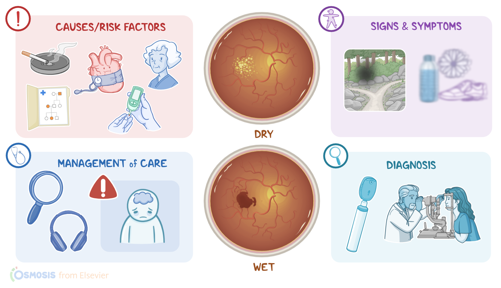
Macular degeneration is an eye condition in which the part of the retina that is responsible for clear vision, called the macula, degenerates. This causes blurred or reduced central vision, and is typically related to aging. There are two main types of macular degeneration: dry or nonexudative, which is the most common one; and wet or exudative.
Okay, but first, a bit of physiology. If we zoom into the wall of the eye, it is made up of three major layers. There's a fibrous outer layer, which contains the cornea and sclera, and helps shield excess light. The middle vascular layer is called the uvea and consists of the iris, pupil, ciliary body, and choroid. Finally, the neural layer consists of the retina, with its own outer pigmented layer, and an inner neural layer that’s composed of photoreceptor cells, called rods and cones, which convert light into neural signals that travel via the optic nerve to the brain for visual processing.
Now, there’s an oval spot in the middle of the posterior retina, called the macula, which contains the highest concentration of cones and is the part of the retina that offers the highest visual acuity.
Alright, now, even though the exact cause of macular degeneration is still unknown, several risk factors have been identified. Modifiable risk factors include smoking, diabetes, hypertension, and hyperlipidemia, whereas non-modifiable risk factors include aging and a family history of macular degeneration.
Now, let’s look at the pathology of macular degeneration. Dry macular degeneration typically develops slowly over time and is characterized by yellowish extracellular deposits of waste materials, known as drusen, that build up between the choroid and the retinal pigment epithelium. On the other hand, in wet macular degeneration, which develops rapidly, there is abnormal neovascularization, meaning that abnormal blood vessels grow from the choroid behind the retina and can leak intravascular fluid or blood.
In terms of signs and symptoms, clients with macular degeneration typically present with blurry and distorted vision, poor night vision, as well as scotomas or missing areas of vision. Eventually, clients can develop central vision loss and become legally blind, even though their peripheral vision remains intact.
The diagnosis of macular degeneration starts with the client’s history and physical assessment, followed by ophthalmoscopy and slit lamp biomicroscopy to examine the retina. In dry macular degeneration, this shows the drusen, whereas in wet macular degeneration, retinal bleeding, and edema can be seen.
Other diagnostic tests include the Amsler grid test, which is basically a grid that has a small central dot and might help better define the areas of the visual field that are missing. In addition, optical coherence tomography can be used to take cross-section pictures of the retina. Fluorescein angiography can be used to show the newly formed abnormal blood vessels in wet macular degeneration.
Now, treatment varies depending on the type of macular degeneration. For dry macular degeneration there’s no cure, so treatment focuses on slowing down vision loss. Maintaining a healthy diet, as well as multivitamin and antioxidant supplements has been shown to help. On the other hand, wet macular degeneration can be treated with intraocular injection of anti-VEGF medications like ranibizumab, that inhibit the formation of new blood vessels, or ocular photodynamic therapy along with a light-sensitive drug called verteporfin. Finally, laser photocoagulation therapy can be also done to seal off the leaking blood vessels.
Alright, let’s take a look at the nursing care you will provide for a client with macular degeneration. Priority nursing goals include promoting independence and safety despite decreased visual acuity, and to support the client with the psychological challenges that come with progressive vision loss.