Myocarditis: Nursing
Myocarditis: Nursing
RN MS Cardiac/Perfusion
RN MS Cardiac/Perfusion
Notes
| MYOCARDITIS | ||
| KEY POINTS | NOTES | |
| DEFINITION |
| |
| PHYSIOLOGY |
| |
| CAUSES AND RISK FACTORS |
| |
| PATHOPHYSIOLOGY |
| |
| SIGNS AND SYMPTOMS |
| |
| DIAGNOSIS |
| |
| TREATMENT |
| |
| MANAGEMENT OF CARE |
| |
| PATIENT AND FAMILY TEACHING |
| |
Transcript
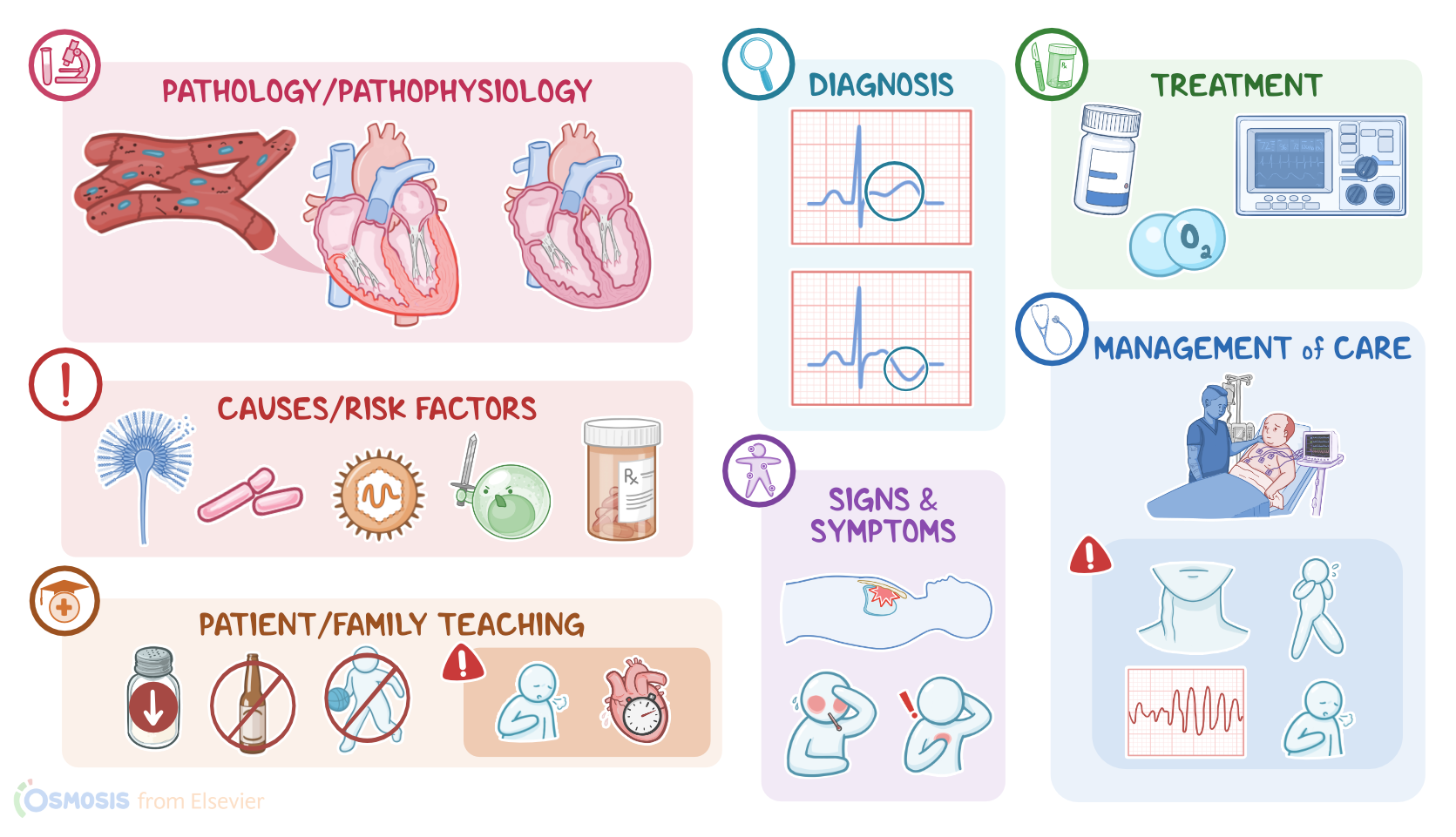
Myocarditis refers to the inflammation of the heart muscle, called the myocardium.
Okay, but first, a bit of anatomy and physiology. The heart wall is made of three layers: the outer layer is the epicardium, the middle layer is the myocardium, and the inner layer is the endocardium.
So, the myocardium is a muscular layer that consists of cells called cardiomyocytes, which relax so that the heart can be filled with blood during diastole, and then contract to pump the blood out of the heart during systole.
Contraction of the cardiomyocytes is controlled by the pacemaker cells in the sinoatrial or SA node, which generates electrical signals that get sent out through the conduction system in the heart.
Now, most cases of myocarditis are caused by a viral infection, such as Coxsackie A or B virus and parvovirus B19; but can also be caused by a bacterial infection, like Streptococcus pyogenes or Borrelia burgdorferi, which causes Lyme disease; a fungal infection, such as Candida or Aspergillus; or a parasitic infection, like Trypanosoma cruzi, which causes Chagas disease.
Myocarditis can also be caused by autoimmune disease, such as systemic lupus erythematosus or polymyositis, where the immune system attacks its own tissues, including the myocardium.
Other causes of myocarditis include toxins like carbon monoxide poisoning, radiation therapy in the chest, and certain medications like sulfonamides, or chemotherapeutic medications, such as anthracycline and doxorubicin. Finally, some cases of myocarditis are idiopathic, meaning the cause is unknown.
Okay, regardless of the cause, the pathological outcome is acute damage to the cardiomyocytes. This in turn activates the immune system, causing the release of cytokines and oxygen free radicals. This results in severe inflammation, and even cardiomyocyte death, which impairs the heart’s ability to contract. Once the causative agent gets cleared out, the inflammation typically resolves, and myocardial contraction returns to normal.
As far as complications go, if myocarditis is severe enough, the inflammation may result in myocardial fibrosis, which can lead to long term problems with contraction and arrhythmias, and even sudden cardiac death Another complication occurs if the body’s immune response remains active, and actually starts attacking the cardiomyocytes. Over time, this can lead to heart dysfunction, and more specifically, to the development of dilated cardiomyopathy, where all four chambers of the heart dilate, while the heart walls become thinner, so contraction becomes weaker, potentially leading to heart failure. Ultimately, some clients may develop life-threatening complications like cardiogenic shock.
Initially, clients with myocarditis may experience flu-like symptoms, such as fever, fatigue, sore throat, malaise, myalgias, and arthralgias. Some clients can also develop dyspnea and lymphadenopathy, as well as nausea and vomiting. These clinical manifestations might be followed by arrhythmias.
If there’s involvement of the pericardium surrounding the heart, clients can also experience chest pain that’s typically positional, meaning that it can get worse when the client is in supine position and with inspiration; and gets better when the client is sitting upright and leaning forward better.
In severe cases, myocarditis may develop into heart failure, leading to fatigue, dyspnea, and palpitations, as well as jugular venous distention, hepatomegaly, and peripheral edema.
Diagnosis of myocarditis starts with history and physical assessment. On electrocardiogram or ECG, clients may have T-wave inversions and “saddle-shaped” ST-segment elevations, as well as sinus tachycardia, or arrhythmias like atrioventricular block.
Laboratory tests might show elevated cardiac enzymes like troponin and brain natriuretic peptide or BNP; as well as leukocytosis; and increased inflammatory markers like CRP and ESR. In the case of viral infection, viral titers might also be raised.
Imaging techniques like echocardiography, nuclear scans, and MRI are sometimes also used to assess the heart function. If necessary, the diagnosis can be confirmed with a biopsy of the myocardium in the first 6 weeks of the acute illness.