Paget disease of bone: Nursing
Introduction0:00–0:27
Paget disease of bone is a disorder in which there’s a lot of bone remodeling that happens in some regions of the bone. Typically, there’s excessive bone resorption followed by disorganized and excessive bone growth, leading to skeletal deformities, fragile bones, and potential fractures.
Paget disease of bone can affect a single bone or several bones, and most often, it involves the skull, spine, pelvis, arms, and legs.Now, let’s quickly review the physiology of bones.Normally, the surface of the bones is covered by a dense layer of connective tissue called the periosteum, and it's where the muscles, tendons, and ligaments are attached.
Physiology0:27–1:29
Beneath the periosteum, there’s a dense and tough external layer called compact bone or cortical bone composed of collagen and hydroxyapatite which contains calcium and phosphate.
In the center of the bone, there’s the medullary canal, a hollow space lined by a honeycomb- looking structure called the spongy or cancellous bone.
The spaces in the spongy bone are occupied by the bone marrow, which is the site of blood cell production.At first glance, a bone may appear inert and unchanging, but it’s actually a very dynamic tissue.
In general, a bone is replaced with new cells every three to ten years in a process called bone remodeling, which has two steps: bone resorption, when specialized cells called osteoclasts break down bone, and bone formation, which is when another type of cells called osteoblasts form new bone.
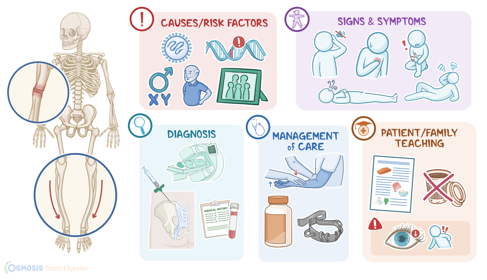
The exact cause of Paget disease of bone is unclear, with some theories suggesting that it can get triggered by infections like the measles virus, and is linked to genetic mutations.
Causes & risk factors1:29–1:49
Risk factors include being assigned male at birth, age over 40, family history of Paget disease, Caucasian race, or northern European descent.
The pathology of Paget disease has three phases, called lytic, mixed, and sclerotic phases. Phase one is the lytic phase, and that’s where osteoclasts increase in number and activity to aggressively demineralize the bone.
Pathology1:49–3:14
Phase two is the mixed phase where the lytic phase continues but the blastic phase also starts. In the blastic phase, there’s a rapid but disorganized proliferation of new bone tissue by an increased number of osteoblasts.
The result is that collagen fibers are deposited in a haphazard way.Phase three is the sclerotic phase, and that’s where new bone formation exceeds bone resorption, and the net result is more bony matter, but the bones are structurally disorganized and therefore weaker than healthy bones.
At the same time, osteoclasts secrete molecules that induce new blood vessel formation, resulting in an increased vascularization of the sclerotic bone.
Eventually, the osteoblastic activity also slows down, and there’s a dormant state in the disease, called the “burned-out state.” Finally, in rare cases, the accelerated bony growth can lead to genetic mutations that cause a type of bone cancer called osteosarcoma.
When that happens it’s referred to as Paget sarcoma. Clinically, clients with Paget disease often have no symptoms.
But when symptoms are present, they can experience fragility fractures, where the bones are so weak that they break with minimal force, like bumping into a door, or even spontaneously from the weight of the body alone.
Clinical manifestations3:14–5:14
Over time, clients can also develop bone pain that ranges from mild to severe, as well as bone deformities, such as bowing of the legs.
Paget disease of bone can also cause neurological symptoms. Misshapen and overgrown bones in the vertebral column can impinge on the spinal nerve roots.
In severe cases, there can also be compression of the cauda equina, which is the bundle of nerves protruding from the bottom of the spinal cord that resembles a horse's tail.
This is a rare but dangerous condition where the client will experience severe lower back and leg pain, weakness and sensation loss in the lower extremities, especially the “saddle region” which includes the inner thigh, groin and buttocks and loss of bladder and bowel control.
Bone overgrowth can also narrow the auditory foramen or optic canal and impinge on the auditory nerve or optic nerve respectively.
This can cause hearing or vision loss, respectively. Next, if the parts of the bone that are involved in a joint are involved, it can result in osteoarthritis.
This can cause joint pain and stiffness. If the skull is involved, the overgrowth of bones can cause headache.
It could also change the shape of the client’s face which is sometimes described as a “ lion- like” appearance and this is called leontiasis.
In addition, increased vascularization of the new bone means that the heart has to work harder to keep up with the increased blood supply.
In severe cases, this can progress to heart failure, which can cause manifestations like orthopnea. Another cardiovascular complication of Paget disease of bone is calcification of the aortic valve, which can lead to aortic stenosis and cause manifestations like dyspnea, particularly during exercise; fatigue; and syncope.
The diagnosis of Paget disease of bone starts with the client's history and physical assessment, followed by blood tests, which typically reveal elevated levels of alkaline phosphatase, which gets released into the blood as bone is destroyed, with normal calcium and phosphate levels.
Diagnosis5:14–6:48
Urinalysis can show increased markers of collagen degradation, especially during the lytic and mixed phases. In addition, an X-ray might show lytic lesions during the lytic phase of the disease, and sclerotic lesions which are thickened and dense areas that develop over time.
A CT or MRI scan, as well as a bone scintigraphy can also be done to identify lesions. Finally, a bone biopsy is usually done to exclude malignancies which can mimic some of the findings of Paget disease of bone.Now, the treatment of mild forms of Paget disease of bone can rely on monitoring the client’s condition, if they are asymptomatic and don’t have any significant laboratory or imaging changes .
Otherwise, treatment is focused mainly on pain management, with NSAIDs or acetaminophen, and trying to limit bone resorption with bisphosphonates like zoledronic acid, risedronate or alendronate.
For clients who can’t tolerate bisphosphonates, subcutaneous injections of calcitonin can be given. Calcium and vitamin D prescriptions are sometimes also administered to correct hypercalcemia before starting treatment with bisphosphonates.
Finally, surgery can help correct bone deformities, decompress an impinged nerve, and reduce associated fractures.Alright, let’s look at the nursing care you’ll be providing for a client with Paget disease.
For most clients, your goals of care are to maintain safety, promote musculoskeletal function, and manage pain.Begin by assessing your client's mobility level, strength, and gait, and placing them on fall precautions.
Management and care6:48–7:41
Provide them with appropriate assistive devices as needed, such as a walker or cane. Use a gait belt when transferring them and when assisting with ambulation.
If braces are recommended, assist them with application. Lastly, collaborate with the physical therapist to promote muscle-strengthening and balance.Next, administer your client's prescribed medications, including a bisphosphonate, calcium and vitamin D, as well as analgesics to reach their pain goal.
Okay, moving on to client and family education. Begin by explaining how Paget disease causes abnormal breakdown and rebuilding of the bones that eventually makes them weaker and more prone to fracture.
General client and family teaching7:41–10:01
Let them know that the plan of care is focused on slowing the abnormal bone growth, which will help prevent fractures, decrease pain, and improve their ability to accomplish their activities of daily living.
Review their prescribed medications, and instruct them to take them exactly as directed. Lastly, remind them of the importance of keeping all their follow-up appointments, including regular laboratory measurements of their serum alkaline phosphatase.Next, review health promotion activities to incorporate into their daily routine to help promote bone health.
Remind them that keeping as active as possible helps to slow bone demineralization, but to avoid aggressive physical activity or contact sports to decrease the risk of fracture.
Discuss the importance of good body mechanics, and stress the importance of avoiding activities such as heavy lifting or twisting that can put undue stress on their bones.
Recommend a diet high in nutrients that are important for healthy bones, including in protein, vitamin D, and calcium. Provide them a list of foods high in these nutrients that they can choose from, and talk to them about how to incorporate them into their diet each day.
Remind them to stay well hydrated by drinking about 2 liters of water each day, while avoiding caffeinated beverages and alcohol.
Reinforce the importance of using assistive devices such as a walker or cane to assist when walking. Finally, instruct them to contact their healthcare provider right away if they experience any new symptoms like changes in vision or hearing, or worsening bone pain.
If they fall or think they might have a broken bone, instruct them to get medical assistance immediately.Alright, as a quick recap… Paget disease of bone is a chronic skeletal bone disorder where excessive bone resorption is followed by disorganized and excessive bone growth, skeletal deformities, fragile bones, and potential fractures.
Review10:01–11:47
Paget disease of bone usually involves the skull, spine, pelvis, arms, and legs. The cause of Paget disease is unknown, but may have viral or genetic factors.
Paget disease is composed of three phases: the lytic phase, where osteoclasts start to aggressively demineralize the bone; the mixed phase, where the lytic phase continues along with a blastic phase, that involves rapid but disorganized proliferation of new bone tissue; and the sclerotic phase, where new bone is course and fibrous.
Complications of Paget disease can include bone deformity, pathologic fractures, nerve impingement, cauda equina syndrome, vision or hearing loss, cardiac complications, or osteosarcoma.Symptoms include bone pain, pathologic fracture, musculoskeletal deformities, joint pain and stiffness, fatigue, skull enlargement, and spinal cord or nerve root compression.
Paget disease is diagnosed in clinical evaluation with a detailed client history, blood tests including alkaline phosphatase, X-ray, as well as CT, MRI and scintigraphic bone scans.Treatment is focused on pain management and preventing bone resorption with medications such as bisphosphonates and calcitonin.
Surgery may also be an option to correct deformities or decompress an impinged nerve.Nursing goals when caring for a client with Paget disease include maintaining safety, promoting musculoskeletal function, and managing pain.
Client and family education is focused on health promotion and maintenance, and safety measures to prevent fractures. health promotion and maintenance and safety measures
| PAGET DISEASE OF BONE | ||
| KEY POINTS | NOTES | |
| DEFINITION |
| |
| PHYSIOLOGY |
| |
| CAUSES AND RISK FACTORS |
| |
| PATHOPHYSIOLOGY |
| |
| SIGNS AND SYMPTOMS |
| |
| DIAGNOSIS |
| |
| TREATMENT |
| |
| MANAGEMENT OF CARE |
| |
| PATIENT AND FAMILY TEACHING |
| |
No notes for this video yet
Try adding a note below