Shock - Neurogenic: Nursing

1,298views

Shock - Neurogenic: Nursing

Watch later

Watch later

Hypokalemia: Clinical
Movement of water between body compartments
The role of the kidney in acid-base balance
Blood histology
Blood components
Respiratory acidosis
Metabolic acidosis
Respiratory alkalosis
Metabolic alkalosis
Metabolic and respiratory alkalosis: Clinical
Metabolic and respiratory acidosis: Clinical
Bronchodilators: Beta 2-agonists and muscarinic antagonists
Chronic obstructive pulmonary disease (COPD): Clinical
Chronic obstructive pulmonary disease (COPD): Nursing process (ADPIE)
Adrenergic antagonists: Alpha blockers
Sympatholytics: Alpha-2 agonists
Alpha-2 adrenergic agonists: Nursing pharmacology
Adrenergic receptors
Adrenergic antagonists: Beta blockers
Diabetes mellitus: Clinical
Diabetes mellitus (DM): Nursing process (ADPIE)
Diabetes mellitus
Diabetes mellitus: Pathology review
Stroke: Nursing process (ADPIE)
Stroke: Clinical
Ischemic stroke
Intracerebral hemorrhage
Peptic ulcers and stomach cancer: Clinical
GERD, peptic ulcers, gastritis, and stomach cancer: Pathology review
Sickle cell disease (NORD)
Sickle cell disease: Nursing process (ADPIE)
Sickle cell disease: Clinical
Blood groups and transfusions
Erythropoietin
Blood products: Nursing pharmacology
Oxygen binding capacity and oxygen content
Blood products and transfusion: Clinical
Hemophilia
Hemophilia: Nursing process (ADPIE)
Leukemia: Nursing process (ADPIE)
Chronic leukemia
Leukemias: Pathology review
Acute leukemia
Leukemia: Clinical
Lymphomas: Pathology review
Lymphoma: Clinical
Non-Hodgkin lymphoma
Hodgkin lymphoma
Lymphatic system anatomy and physiology
Anatomy of the abdominal viscera: Liver, biliary ducts and gallbladder
Liver anatomy and physiology
Cirrhosis
Cirrhosis: Pathology review
Cirrhosis: Clinical
Cirrhosis: Nursing process (ADPIE)
Hepatitis C virus
Hepatitis A and Hepatitis E virus
Liver cancer: Nursing
Cholestatic liver disease
Non-alcoholic fatty liver disease
HIV (AIDS)
HIV and AIDS: Pathology review
Antiretrovirals for HIV/AIDS - Protease inhibitors: Nursing pharmacology
Antiretrovirals for HIV/AIDS - NRTIs and NNRTIs: Nursing pharmacology
Antiretrovirals for HIV/AIDS - Integrase strand transfer inhibitors: Nursing pharmacology
Anatomy of the abdominal viscera: Pancreas and spleen
Pancreatitis: Nursing process (ADPIE)
Glaucoma: Nursing process (ADPIE)
Glaucoma
Eye conditions: Refractive errors, lens disorders and glaucoma: Pathology review
Acute respiratory distress syndrome (ARDS): Nursing
Anatomy of the coronary circulation
Coronary artery disease: Pathology review
Coronary artery disease: Clinical
ECG basics
ECG normal sinus rhythm
ECG rate and rhythm
ECG axis
ECG intervals
ECG QRS transition
ECG cardiac infarction and ischemia
Cardiac cycle
Arterial disease
Delirium
Dementia and delirium: Clinical
Vascular dementia
Frontotemporal dementia
Dementia with Lewy bodies
Dementia: Pathology review
Tumor lysis syndrome (TLS): Nursing Process (ADPIE)
Cholelithiasis: Nursing
Coronary artery disease (CAD) and angina pectoris: Nursing process (ADPIE)
Breast cancer: Nursing process (ADPIE)
Ovarian cancer: Nursing
Cervical cancer: Nursing
Hormones and hormone modulators for cancer: Nursing pharmacology
Endometriosis: Nursing
Heart failure
Left-sided heart failure: Nursing process (ADPIE)
Heart failure: Pathology review
Pneumonia: Pathology review
Pneumonia
Bacterial pneumonia: Nursing process (ADPIE)
Respiratory stimulants: Nursing pharmacology
Corticosteroids - Inhaled: Nursing pharmacology
Pneumothorax and hemothorax: Nursing
Chest trauma: Clinical
Pleural effusion: Nursing
Tuberculosis (TB): Nursing
Parkinson disease: Nursing process (ADPIE)
Huntington disease
Multiple sclerosis (MS): Nursing
Multiple sclerosis
Guillain-Barré syndrome: Nursing
Guillain-Barre syndrome
Myasthenia gravis: Nursing
Acute kidney injury (AKI): Nursing process (ADPIE)
Acute kidney injury: Clinical
Chronic kidney disease (CKD): Nursing
Chronic kidney disease
Chronic kidney disease: Clinical
Polycystic kidney disease (PKD): Nursing
Renin-angiotensin-aldosterone system
Osteoarthritis: Nursing
Osteoarthritis
Rheumatoid arthritis (RA): Nursing process (ADPIE)
Rheumatoid arthritis: Clinical
Rheumatoid arthritis
Rheumatoid arthritis and osteoarthritis: Pathology review
Systemic lupus erythematosus
Systemic lupus erythematosus (SLE): Pathology review
Systemic lupus erythematosus (SLE): Clinical
Systemic lupus erythematosus (SLE): Nursing
Mycobacterium tuberculosis (Tuberculosis)
Peripheral venous disease (PVD): Nursing process (ADPIE)
Peripheral arterial disease (PAD): Nursing process (ADPIE)
Buerger disease: Nursing
Raynaud phenomenon: Nursing
Aortic dissections and aneurysms: Pathology review
Aortic aneurysms and dissections: Clinical
Aortic dissection
Aortic aneurysm: Nursing process (ADPIE)
Venous thromboembolism (VTE): Nursing process (ADPIE)
Benign prostatic hyperplasia (BPH): Nursing process (ADPIE)
Benign prostatic hyperplasia
Prostate cancer: Nursing
Prostate cancer
Testicular cancer: Nursing
Testicular cancer
Diabetic ketoacidosis (DKA): Nursing process (ADPIE)
Endocrine system anatomy and physiology
Vitamin B12 deficiency
Thalassemia: Nursing
Anemia - Iron-deficiency: Nursing
Anemia - Macrocytic: Nursing
Anemia - Aplastic: Nursing
Thyroid hormones
Medications for thyroid disorders: Nursing pharmacology
Hyperthyroidism: Nursing process (ADPIE)
Hypothyroidism: Nursing process (ADPIE)
Hypoparathyroidism: Nursing
Hyperparathyroidism: Nursing
Anxiety disorders: Nursing process (ADPIE)
Spinal cord injury (SCI): Nursing
Cluster A personality disorders
Smoke inhalation injury: Nursing process (ADPIE)
Shock - Anaphylactic: Nursing
Shock - Obstructive: Nursing
Shock - Neurogenic: Nursing
Shock - Hypovolemic: Nursing
Shock - Cardiogenic: Nursing
Shock - Septic: Nursing
Pulmonary edema: Nursing
Burn injury: Nursing

Notes

SHOCK - NEUROGENIC

KEY POINTS
NOTES
DEFINITION
  • Shock: life-threatening condition when organs don't receive enough oxygen and nutrients to function
  • Type of distributive shock
  • Brain or spinal cord damage causes cardiovascular dysfunction

PHYSIOLOGY
  • Blood vessels contain smooth muscle
  • When relaxed, diameter increased
    • Vasodilation
  • When contracted, diameter decreased
    • Vasoconstriction
  • Sympathetic nervous system (SNS) can increase or decrease diameter based on body's needs
    • Increased SNS: vasoconstriction
      • Increased blood pressure
    • Decreased SNS: vasodilation
      • Decreased blood pressure

CAUSES AND RISK FACTORS
  • Causes
    • Trauma to cervical or upper thoracic spinal cord
    • Traumatic brain injury
    • Guillain-Barre syndrome
    • Transverse myelitis
    • Spinal anesthesia
  • Risk factors
    • Any factor that increases risk of trauma to brain or spinal cord
      • High-risk behaviors
      • Bone or joint disorders

PATHOPHYSIOLOGY
  • Damage to SNS neurons
    • PNS goes unbalances
  • Systemic vasodilation
    • Decreased venous return
    • Cardiac output (CO) decreases
  • Body cells switch to anaerobic metabolism
    • Lactic acid builds up
  • Body unable to increase cardiac output
  • Severe tissue hypoxia
  • Multiple organ failure

SIGNS AND SYMPTOMS
  • Hypotension
  • Weak peripheral pulses
  • Altered mental status
  • Coma
  • Skin warm and flush
  • Bradycardia

DIAGNOSIS
  • History
  • Physical assessment
  • X-ray
  • CT
  • MRI

TREATMENT
  • Stabilize spine
  • Secure airway
  • Provide oxygen
  • IV fluids
  • Vasopressors
  • Corticosteroids
  • Address underlying cause

MANAGEMENT OF CARE
  • Goals of care
    • Support ventilation and oxygenation
    • Promote CO
    • Monitor for complications
    • Provide emotional support
  • Apply cervical collar
  • Keep head in neutral position
  • Elevate head of bed
  • Apply high flow oxygen
  • Monitor respiratory status and initiate continuous pulse oximetry
    • Report to HCP
      • Dyspnea
      • Low oxygen saturation
      • Ineffective cough
      • Tachypnea
      • Diminished or crackles lung sounds
  • Administer IV fluids as prescribed
  • Insert indwelling catheter
  • Assess vital signs
    • Report to HCP
      • Signs of decreased CO
  • Assist in preparation for surgery as needed
  • Monitor for complications
    • Report to HCP
      • Signs of autonomic dysreflexia
    • Monitor temperature
    • Prevent skin breakdown and deep vein thrombosis
  • Provide emotional support
  • Coordinate care with interdisciplinary team

PATIENT AND FAMILY TEACHING
  • Explain condition, plan of care, and how to safely self-administer medications
  • Encourage engagement in rehab activities
  • Refer to support groups
  • Instruct on toileting schedule and use of urinary catheter
  • Encourage oral fluids and diet high in fiber
  • Seek emergency care
    • Signs of autonomic dysreflexia
  • Remind to change positions every 2 hours, use devices to distribute pressure, and perform skin care
  • Notify HCP
    • Difficulty with urinary or bowel elimination
    • Sores

Transcript

Watch video only

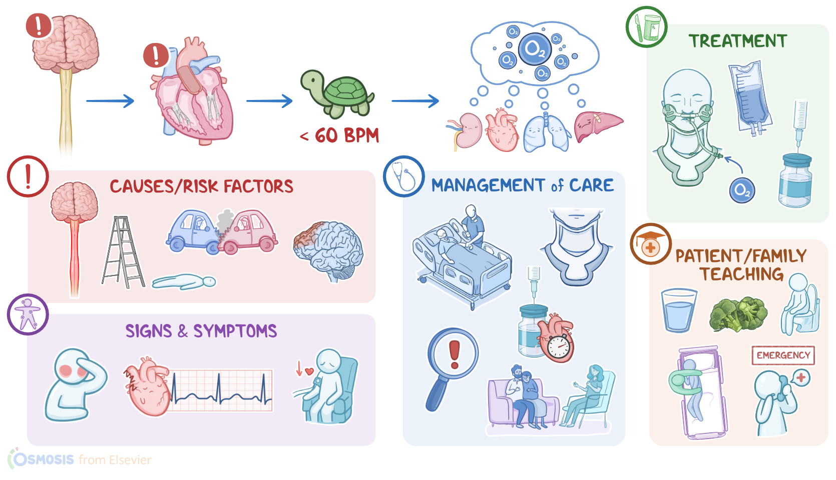
Shock is a life-threatening condition that occurs when the organs don’t receive enough oxygen and nutrients to function properly. Shock can be grouped into four types based on the cause: hypovolemic, cardiogenic, obstructive, and distributive shock. Neurogenic shock is a type of distributive shock where damage to the brain or spinal cord causes dysregulation of the cardiovascular system, leading to decreased heart rate and vasodilation, which eventually leads to impaired tissue perfusion.

Now, to understand the neurogenic shock, let’s quickly review the physiology of blood vessels, which contain smooth muscle in their walls. When the smooth muscle relaxes, it increases the diameter of blood vessels, called vasodilation. On the other hand, when smooth muscle contracts, the diameter of blood vessels decreases, called vasoconstriction.

The contraction and relaxation of smooth muscles are primarily controlled by the sympathetic nervous system, which normally maintains a partial constriction, generating enough force to keep blood moving through the circulatory system. The sympathetic system stimulation can increase or decrease to keep up with the body’s needs. Increased sympathetic stimulation of the blood vessels causes increased heart rate and vasoconstriction, which increases peripheral vascular resistance. Vasoconstriction, when the total blood volume is constant, raises blood pressure and allows blood to flow faster through the blood vessels. In contrast, decreased heart rate and vasodilation is typically caused by decreased sympathetic stimulation, in addition to inflammatory cytokines and histamine, which decrease peripheral vascular resistance. So, vasodilatation, when blood volume is constant, lowers the blood pressure and slows down the blood flow through the blood vessels.

Now let’s switch gears and look at the causes of neurogenic shock. This type of shock is primarily caused by trauma to cervical or upper thoracic segments of the spinal cord, which is typically associated with motor vehicle crashes, as well as severe falls, such as falling from a ladder. Other, less common causes include traumatic brain injury, Guillain-Barre syndrome, transverse myelitis, as well as spinal anesthesia.

Risk factors for developing neurogenic shock include anything increasing the risk of spinal cord trauma, such as engaging in high-risk behaviors, like speeding, and not wearing safety equipment when playing sports. Additionally, clients with a history of bone or joint disorders are also at a higher risk, since even minor trauma could damage the weakened bones, thereby injuring the spinal cord and potentially resulting in neurogenic shock.

The pathology behind neurogenic shock is based on the dysregulation between the sympathetic and parasympathetic stimulation of the cardiovascular system. Injury to the spinal cord can damage the neurons responsible for sympathetic innervation. As a result, the balance is tipped towards the parasympathetic system, causing bradycardia and massive peripheral vasodilation, causing blood to remain in the limbs instead of returning to the heart. All these factors together decrease the blood pressure and organ perfusion.

Now, most types of shock can be divided into four main stages: initial, compensatory, progressive, and refractory. But neurogenic shock deviates from this pattern. Things are pretty similar to other types of distributive shock during the initial stage, when systemic vasodilation causes less blood to return to the heart through the venous system; this, in turn, decreases cardiac output. As a consequence, body cells are not well-perfused, so they switch to anaerobic metabolism to produce energy, causing lactic acid to build up in the blood. When lactic acid builds up, that lowers blood pH, causing metabolic acidosis. Normally, this activates the compensatory stage, which involves stimulation of the sympathetic nervous system to increase the heart rate and vasoconstriction. However, in neurogenic shock, there’s a loss of the sympathetic activity, so the body fails to increase cardiac output. Therefore, with neurogenic shock, there’s no compensatory stage. If not managed in time neurogenic shock can progress to severe tissue hypoxia; and vital organs like the heart, brain, and kidneys may begin to shut down, leading to multiple organ failure.

Clinical manifestations of neurogenic shock share many similarities with other types of shock. There's hypotension, weak peripheral pulse and in severe cases, altered mental status or coma. Now, unlike with hypovolemic, obstructive, or cardiogenic shock, the skin can be warm and flush instead of cold and pale, and this is due to increased peripheral vasodilation. Furthermore, neurogenic shock is the only type of shock where there’s bradycardia instead of tachycardia since sympathetic regulation of the heart is impaired.

Spinal cord injury that causes neurogenic shock is often accompanied by other neurological symptoms. These include muscle weakness or paralysis, urinary incontinence, impaired respiration, and the inability to regulate temperature, where the client becomes poikilothermic, meaning they become dependent on the environment for temperature regulation.

Diagnosis of neurogenic shock starts with the client’s history and physical assessment and it’s made based on the symptoms and ruling out other causes of shock. This is followed by imaging tests like X-rays, CT scan, or MRI to look for the exact cause and location of the spinal cord or brain injury.

Finally, let’s take a look at treatment. The first step in the management of the client with neurogenic shock is to stabilize the client’s spine using a cervical collar, secure the airway, and provide oxygen. Some clients might also need mechanical ventilatory support. The management continues with early resuscitation with IV fluids and administration of vasoactive medications like norepinephrine. In the case of persistent bradycardia, atropine can be used to increase the heart rate. Finally, clients with neurogenic shock might benefit from the administration of corticosteroids, such as methylprednisolone. Finally, it’s important to address the underlying cause whenever possible, like performing spinal decompression procedures such as closed reduction using traction, or spinal surgery.